1
BiP | Lumenal-binding protein (chicken antibody)
AS09 614 | Clonality: Polyclonal | Host: Hen | Reactivity: Arabidopsis thaliana, Hordeum vulgare, Spinacia oleracea, Zea mays
Limited stock
- Product Info
-
Immunogen: KLH-conjugated synthetic peptide derived from Arabidopsis thaliana BiP proteins: BiP1 UniProt:Q9LKR3, TAIR: At5g28540, BiP2 UniProt: F4K007, TAIR: At5g42020, BiP3 UniProt:Q8H1B3 ,TAIR:At1g09080
Host: Chicken Clonality: Polyclonal Purity: Immunogen affinity purified total IgY. in PBS pH 7.4. Format: Liquid Quantity: 100 �g Storage: Store at 4�C; once reconstituted make aliquots to avoid repeated freeze-thaw cycles. Please remember to spin the tubes briefly prior to opening them to avoid any losses that might occur from material adhering to the cap or sides of the tube. Tested applications: Immunofluorescence (IF), Western blot (WB) Recommended dilution: 1 : 50-1 : 1000 (IF), 1 : 2000 (WB) Expected | apparent MW: 73,5 | 80 kDa - Reactivity
-
Confirmed reactivity: Arabidopsis thaliana, Hordeum vulgare, Physcomitrium patens, Spinacia oleracea, Zea mays
Predicted reactivity: Nicotiana tabacum, Oryza sativa, Physcomitrium patens, Piea sitchensis, Populus trichocarpa, Spinacia oleracea, Zea mays
Species of your interest not listed? Contact usNot reactive in: No confirmed exceptions from predicted reactivity are currently known - Application Examples
-
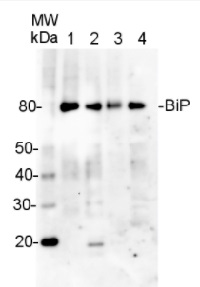
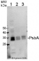
Application example

5 µg of total protein from A.thaliana (1), H. vulgare (2), Z.mays (3), S. oleracea (4), extracted with Agrisera PEB extraction buffer (AS08 300) were separated on 4-12% SDS-PAGE and blotted 1h to PVDF. Blots were blocked immediately following transfer in for 1h at room temperature with agitation. Blots were incubated in the primary antibody at a dilution of 1: 10 000 for 1h at room temperature with agitation. The antibody solution was decanted and the blot was rinsed briefly twice, then washed once for 15 min and 3 times for 5 min in TBS-T at room temperature with agitation. Blots were incubated in secondary antibody (anti-hen IgY horse radish peroxidase conjugated, from Agrisera AS09 603) diluted to 1:50 000 for 1h at room temperature with agitation. The blots were washed as above and developed for 5 min with ECL detection reagent according to the manufacturers instructions. Exposure time was 5 seconds.
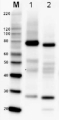
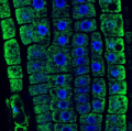
immunofluorescence

BiP localization in 5 days old Arabidopsis thaliana roots. BiP signal shown in green, DAPI in blue. The material has been fixed in para-formaldehyde for 30 minutes. Tissue cleaning has been performed before immunolocalization. Chicken anti-BiP primary antibody was diluted in 1: 1000 and DyLight®488 conjugated goat anti-chicken secondary antibody AS09 622 (green color) was diluted in 1: 1000. Co-staining with DAPI visualized nucleus (blue color). Scale bar – 10 µm.
Courtesy Dr. Taras Pasternak, Freiburg University, Germany
- Additional Information
-
Additional information: Antibody solution contains 0,02% sodium azide as preservative Additional information (application): Protein or membrane sample should be treated at 70�C for 10 min before loading on the gel - Background
-
Background: BiP2 (Binding immunoglobulin protein) is localized in endoplasmic reticulum lumen (ER) and plays a role in protein assembly inside ER. BiP protein is abundant under all growth conditions but its synthesis can increase under conditions that lead to the accumulation of unfolded polypeptides in endoplasmic reticulum (ER). Alternative name: AtBP2
- Product Citations
-
Selected references: Bennett et al. (2014). Plasma Membrane-Targeted PIN Proteins Drive Shoot Development in a Moss. Curr Biol. 2014 Dec 1;24(23):2776-85. doi: 10.1016/j.cub.2014.09.054. Epub 2014 Nov 13.
- Protocols
-
Agrisera Western Blot protocol and video tutorials
Protocols to work with plant and algal protein extracts
Oxygenic photosynthesis poster by prof. Govindjee and Dr. Shevela
Z-scheme of photosynthetic electron transport by prof. Govindjee and Dr. Björn and Dr. ShevelaMethod of isolation of plant ER
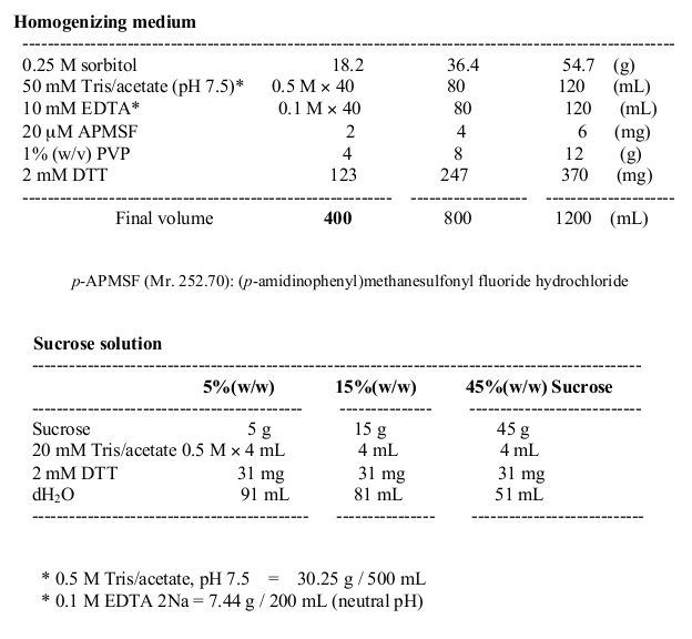
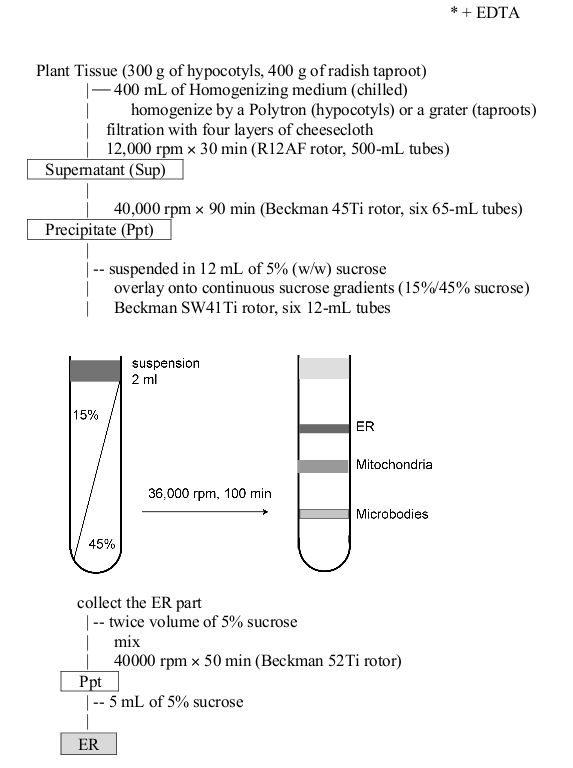
Courtesy of Dr. Masayoshi Maeshima
- Reviews:
-
This product doesn't have any reviews.
Accessories
AS09 481 | Clonality: Polyclonal | Host: Rabbit | Reactivity: A. thaliana, B. napus, Chara australis, C. reinhardtii, C. sativus, M. perniciosa, N. benthamiana, N. tabacum, R. sativa, L.Tokinashi-daikon, O. europaea, O. sativa, P. abies, P. patens, S. oleracea, S. lycopersicum, S. tuberosum, T. aestivum, Z. mays
AS09 622 | Clonality: Polyclonal | Host: Goat | Reactivity: Chicken IgY (H&L)
AS09 603 | Clonality: Polyclonal | Host: Goat | Reactivity: Hen IgY (H&L)