1
Anti-Ramy 3D | Alpha-amylase isozyme 3D
AS10 716 | Clonality: Polyclonal | Host: Rabbit | Reactivity: Oryza sativa
- Product Info
-
Immunogen: KLH-conjugated synthetic peptide derived from known Oryza sativa P27933
Host: Rabbit Clonality: Polyclonal Purity: Immunogen affinity purified serum in PBS pH 7.4. Format: Lyophilized Quantity: 100 µg Reconstitution: For reconstitution add 100 µl of sterile water Storage: Store lyophilized/reconstituted at -20°C; once reconstituted make aliquots to avoid repeated freeze-thaw cycles. Please remember to spin the tubes briefly prior to opening them to avoid any losses that might occur from material adhering to the cap or sides of the tube. Tested applications: Western blot (WB) Recommended dilution: 1 : 5000 (WB) Expected | apparent MW: 50 kDa
- Reactivity
-
Confirmed reactivity: Oryza sativa Not reactive in: No confirmed exceptions from predicted reactivity are currently known - Application Examples
-
Application example 
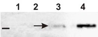
25 µg of total protein from Oryza sativa seedlings (from 1 day to 5 days of imbibition on filter paper at 28°C) extracted with an SDS Extraction Buffer (60mM Tris-HCl pH 8.0, 2% SDS, 1,5% Sucrose) were separated on XT CRITERION 10%Bis-Tris (BioRad) SDS-PAGE and blotted 1h to PVDF. Blot was blocked immediately in milk in TBS-T for 1h at room temperature (RT) with agitation. Blot was incubated in the primary antibody at a diluition of 1: 5000 in milk in TBS-T for 3h at RT with agitation. The antibody solution was decanted and the blot was rinsed briefly once, then washed once for 15 min and 3 times for 5 min in TBS-T at RT with agitation. Blot was incubated in secondary antibody (goat anti-rabbit IgG HRP conjugated, AS09 602) diluited 1:20 000 in milk in TBS-T for 50 min at RT and then washed as above and developed for 3 min with standard ECL. Images of the blot were obtained using BioSpectrum AC Imaging System (UVP). Exposure time was 30 min. The arrow indicates RAmy3D (around 50KDa).
Courtesy Valeria Banti and prof. Pierodomenico Perata, PlantLab, Scuola Superiore Sant'Anna, Italy

25 µg of total protein from Oryza sativa embryos and aleurones (espectively E and A in the figure) were treated with Glucose 100mM and GA3 for 24 hr and 48 hr. Then protein were extracted with an SDS Extraction Buffer (60mM Tris-HCl pH 8.0, 2% SDS, 1,5% Sucrose) and were separated on XT CRITERION 10%Bis-Tris (BioRad) SDS-PAGE and blotted 1h to PVDF. Blot was blocked immediately in milk in TBS-T for 1h at room temperature (RT) with agitation. Blot was incubated in the primary antibody at a diluition of 1: 5000 in milk in TBS-T for 3h at RT with agitation. The antibody solution was decanted and the blot was rinsed briefly once, then washed once for 15 min and 3 times for 5 min in TBS-T at RT with agitation. Blot was incubated in secondary antibody (goat anti-rabbit IgG HRP conjugated, AS09 602) diluited 1:20 000 in milk in TBS-T for 1 hr at RT and then washed as above and developed for 3 min with standard ECL. Images of the blot were obtained using BioSpectrum AC Imaging System (UVP). Exposure time was 30 min. The arrow indicates RAmy3D (around 50KDa), present in embryos at 24 hr and 48hr and completely repressed by glucose.
RAmy3D is expressed in the scutellar epithelium, is mainly under metabolic (sugar) control, with hormones playing little if any role (Karrer and Rodriguez, 1992,Plant Journal; Perata et al., 1997, The Plant Cell). We have confirmed this physiological mechanism treating different tissues (embryo and aleurone+endosperm) with inhibiting agent (glucose 100mM) and GAs (Gibberellic acid). The result (Fig.3) clearly shows the down-regulation by sugars (no signal was detected in presence of glucose), whereas GAs have only a slightly or no effect on Ramy3D.
Courtesy Valeria Banti and prof. Pierodomenico Perata, PlantLab, Scuola Superiore Sant'Anna, Italy
- Background
-
Background: α-Amylases are hydrolytic enzymes responsible for the mobilization of the starch into metabolizable sugars. This process provides the energy for the growth of roots and shoots and is crucial during germination of cereal seeds. These enzymes are coded by a multigene family and even thought other amylolytic enzyme participate in the process of starch breakdown, the contribution of α-amylase is the prerequisite for the initiation of this process. Ramy3D is one of the alpha amylases genes in the rice multigene family (Huang et al. Nucleic Acid Research 1990).
Synonymes:1,4-alpha-D-glucan glucanohydrolase
- Product Citations
-
Selected references: Ye et al. (2018). Natural variation in the promoter of rice calcineurin B-like protein10 (OsCBL10) affects flooding tolerance during seed germination among rice subspecies. Plant J. 2018 May;94(4):612-625. doi: 10.1111/tpj.13881.
Ho et al. (2017). A calcineurin B-like protein participates in low oxygen signalling in rice. CSIRO PUBLISHING Functional Plant Biology. - Protocols
- Agrisera Western Blot protocol and video tutorials
- Reviews:
-
This product doesn't have any reviews.
Related products

AS09 602 | Clonality: Polyclonal | Host: Goat | Reactivity: Rabbit IgG (H&L)

AS09 607 | Clonality: Polyclonal Host: Goat Reactivity: Rabbit IgG (H&L)