1
Anti-CDC2 | Cell-division-cycle kinase 2
AS06 153 | Clonality: Polyclonal | Host: Rabbit | Reactivity: A. thaliana, H. vulgare, O. sativa, P. yunnaniensis, Z. mays, V.faba
- Product Info
-
Immunogen: KLH-conjugated synthetic peptide derived form Zea mays CDC2 sequence P23111, well conserved in other plant species
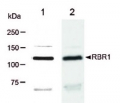
Host: Rabbit Clonality: Polyclonal Purity: Serum Format: Lyophilized Quantity: 50 µl Reconstitution: For reconstitution add 50 µl of sterile water Storage: Store lyophilized/reconstituted at -20°C; once reconstituted make aliquots to avoid repeated freeze-thaw cycles. Please remember to spin the tubes briefly prior to opening them to avoid any losses that might occur from material adhering to the cap or sides of the tube. Tested applications: Immunolocalization (IL), Western blot (WB) Recommended dilution: 1 : 500 (IL), 1 : 2000-1 : 10 000 (WB) Expected | apparent MW: 34-36 (Zea mays)
- Reactivity
-
Confirmed reactivity: Arabidopsis thaliana, Hordeum vulgare,Vicia faba, Oryza sativa, Pinus yunnanensi, Zea mays Predicted reactivity: Pisum sativum, Solanum lycopersicum, Triticum aestivum, Pinus contorta, Populus balsamifera, Physcomitrium patens Not reactive in: No confirmed exceptions from predicted reactivity are currently known - Application Examples
-
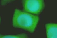
Application example

Seeds of field bean (Vicia faba L. subsp. minor var. Nadwiślański; DANKO Group; Sobiejuchy) were sterilized
using sodium hypochlorite (0.3% v/v) and germinated in Petri dishes on wetted filter paper at room
temperature. At 4 d after imbibition, dark-grown seedlings with primary roots 25±5 mm long were selected for experiments. During incubations roots were oriented horizontally in a humid chamber and aerated continuously on a rotary water-bath shaker (30 rpm) at 23°C.
Immunocytochemical assays were performed according to the method prescribed earlier (Rybaczek and
Maszewski 2006). Excised apical parts of roots (1.5 mm long) were fixed for 45 min (18°C) in PBS-buffered
3.7% paraformaldehyde, washed several times with PBS and placed in a citric acid-buffered digestion solution (pH 5.0; 37°C for 45 min) containing 2.5% pectinase (Fluka), 2.5% cellulase (Onozuka R-10; Serva) and 2.5% pectoliase (ICN). After removing the digestion solution, root tips were washed 3 times in PBS, rinsed with distilled water and squashed onto Super Frost Plus glass slides (Menzel-Gläser). Air-dried slides were pretreated with PBS-buffered 5% BSA at 20°C for 50 min and incubated overnight in a humidified atmosphere (4°C) with rabbit antibody raised against CDC2 (Agrisera), dissolved in PBS containing 1% BSA (at a dilution of 1:500). Following incubation, slides were washed 3 times with PBS and incubated for 1 h (18°C) with
secondary goat anti-rabbit IgG DyLight®488 antibody (AS09 633, Agrisera; 1:3000). Nuclear DNA was stained with 4’,6-diamidino-2-phenyl-indole (DAPI, 0.4 μg/ml; Sigma-Aldrich). Following washing with PBS, slides were air dried and embedded in Vectashield Mounting Media for Fluorescence (Vector Laboratories). Observations were made using Optiphot-2 fluorescence microscope (Nikon) equipped with B-2A filter (blue light; λ ≈ 495 nm) for DyLight-conjugated antibodies and UV-2A filter (UV light; λ ≈ 365 nm) for DAPI. All images were recorded at exactly the same time of integration using DXM 1200 CCD camera.Courtesy Dr. Dorota Rybaczek, Łódź University, Poland
- Background
-
Background: Programmed cell death (PCD) is a fundamentally important biological process required to maintain the integrity and homeostasis of multicellular organisms during normal development and as a response to adverse environments. Cdc2 kinase, plays a major role in driving the cell cycle. Synonymes (for Arabidopsis thaliana): CDC2A, CDKA-1
- Product Citations
-
Selected references: Syu et al. (2014). Impacts of size and shape of silver nanoparticles on Arabidopsis plant growth and gene expression. Plant Physiology and Biochemistry 2014. In print. - Protocols
-
Agrisera Western Blot protocol and video tutorials
Protocols to work with plant and algal protein extracts - Reviews:
-
This product doesn't have any reviews.
Related products

AS09 602 | Clonality: Polyclonal | Host: Goat | Reactivity: Rabbit IgG (H&L)

AS09 607 | Clonality: Polyclonal Host: Goat Reactivity: Rabbit IgG (H&L)
AS11 1627 | Clonality: Polyclonal | Host: Chicken | Reactivity: Arabiodpis thaliana, Medicago sativa