1
Anti-PsaE | PSI-E subunit of photosystem I (monocot)
AS04 047 | Clonality: Polyclonal | Host: Rabbit | Reactivity: Hordeum vulgare, Oryza sativa
- Product Info
-
Immunogen: 10 kDa PSI-E protein purified from barley thylakoids corresponding to PSI-E protein of Horderum vulgare, UniProt: P13194
Host: Rabbit Clonality: Polyclonal Purity: Total IgG. Protein G purified in PBS pH 7.4. Format: Lyophilized Quantity: 100 µl Reconstitution: For reconstitution add 100 µl of sterile water Storage: Store lyophilized/reconstituted at -20°C; once reconstituted make aliquots to avoid repeated freeze-thaw cycles. Please remember to spin the tubes briefly prior to opening them to avoid any losses that might occur from material adhering to the cap or sides of the tube. Tested applications: Western blot (WB) Recommended dilution: 1 : 2000-1 : 5000 (WB) Expected | apparent MW: 10.8 | 10.5 kDa - Reactivity
-
Confirmed reactivity: Hordeum vulgare, Oryza sativa Predicted reactivity: Catalpa bungei, Nicotiana sylvestris, Zea mays
Species of your interest not listed? Contact usNot reactive in: Arabidopsis thaliana, cyanobacteria, Chlamydomonas reinhardtii, Synechococcus sp. PCC7942
- Application Examples
-
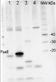
Application example

2 µg of total protein from Arabidopsis thaliana leaf (1), Horderum vulgare leaf (2), Chlamydomonas reinhardtii total cell (3), Synechococcus sp.PCC 7942 total cell (4), all extracted with PEB (AS08 300) were separated on 4-12% NuPage (Invitrogen) LDS-PAGE and blotted 1h to PVDF. Blots were blocked immediately following transfer in 2% blocking reagent in 20 mM Tris, 137 mM sodium chloride pH 7.6 with 0.1% (v/v) Tween-20 (TBS-T) for 1h at room temperature with agitation. Blots were incubated in the primary antibody at a dilution of 1: 10 000 for 1h at room temperature with agitation. The antibody solution was decanted and the blot was rinsed briefly twice, then washed once for 15 min and 3 times for 5 min in TBS-T at room temperature with agitation. Blots were incubated in secondary antibody (anti-rabbit IgG horse radish peroxidase conjugated) diluted to 1:20 000 in 2% blocking solution for 1h at room temperature with agitation. The blots were washed as above and developed for 5 min with chemiluminescence detection reagent according the manufacturers instructions. Images of the blots were obtained using a CCD imager (FluorSMax, Bio-Rad) and Quantity One software (Bio-Rad). - Background
-
Background: PsaE is a nucleus encoded subunit of the Photosystem I reaction center. It is located on the stroma side and interacts with PsaF. PsaE may be involved in Fd reduction. Alternative name: Photosystem I 10.8 kDa polypeptide
- Product Citations
-
Selected references: Yoshida et al. (2016). Hisabori T1.Two distinct redox cascades cooperatively regulate chloroplast functions and sustain plant viability. Proc Natl Acad Sci U S A. 2016 Jul 5;113(27):E3967-76. doi: 10.1073/pnas.1604101113. Epub 2016 Jun 22.
Ye et al. (2012). A Mutation of OSOTP 51 Leads to Impairment of Photosystem I Complex Assembly and Serious Photo-damage in Rice. J Integr Plant Biol. Feb 2012.
Yadavalli et al. (2012). Differential degradation of photosystem I subunits under iron deficiency in rice. J Plant Physiol. March 22. - Protocols
-
Agrisera Western Blot protocol and video tutorials
Protocols to work with plant and algal protein extracts - Reviews:
-
Mathias Pribil | 2011-09-15This antibody works well with Zea mays. PsaE is detectable in mesophyll cell preparations as well as bundle sheath cells There are minor unspecific bands detectable.
Related products

AS09 602 | Clonality: Polyclonal | Host: Goat | Reactivity: Rabbit IgG (H&L)

AS09 607 | Clonality: Polyclonal Host: Goat Reactivity: Rabbit IgG (H&L)