1
Anti-FtsH3 + FtsH10 | ATP-dependent zinc metalloprotease FtsH3 + FtsH10 (mitochondrial)
AS07 204 | Clonality: Polyclonal | Host: Rabbit | Reactivity: Arabidopsis thaliana
- Product Info
-
Immunogen: KLH-conjugated peptide dereived from sequences of Arabidopsis thaliana FtsH3 and FtsH10 with localization to mitochondria Q84WU8, At2g29080 and Q8VZI8, At1g07510
Host: Rabbit Clonality: Polyclonal Purity: Immunogen affinity purified serum in PBS pH 7.4. Format: Lyophilized Quantity: 200 µg Reconstitution: For reconstitution add 100 µl of sterile water Storage: Store lyophilized/reconstituted at -20°C; once reconstituted make aliquots to avoid repeated freeze-thaw cycles. Please remember to spin the tubes briefly prior to opening them to avoid any losses that might occur from material adhering to the cap or sides of the tube. Tested applications: Blue Native PAGE (BN-PAGE), Western blot (WB) Recommended dilution: 1 : 500-1 : 1000 (WB) Expected | apparent MW: 80 kDa
- Reactivity
-
Confirmed reactivity: Arabidopsis thaliana Predicted reactivity: Arabidopsis thaliana
Not reactive in: No confirmed exceptions from predicted reactivity are currently known - Application Examples
-
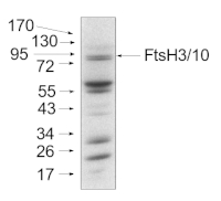
Application example
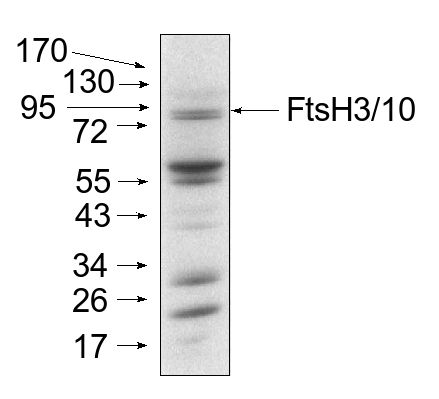
Total protein from Arabidopsis thaliana mitochondria (20 µg) were separated on 10% acrilamide gel and electrophoresis prepared according to Schägger and von Jagov (Anl. Biochem., 1987, 166:368-379). After running the gel, proteins were transferred to nitrocellulose membrane using wet transfer (0.22% CAPS, pH 11). Transfer was checked by Ponceau S staining. Blot was destained by several quick washings in distilled water and 1 washing in 1X TBS (10 mM T pH 7.5, 150 mM NaCl) (10-15 min.).Blot was blocked by 1.5 hour in 5% milk in TBST (1X TBS, 0,1 20) After blocking blot was washed quickly twice in TBST and incubated 2 hours with primary antibody (dilution 1: 1000 TBST (dilution 1:1000). Washing: two quick washings in TBST and 3 x 10 min. washings in TBST. Then blot was incubated 45-60 min. with a secondary anti-rabbit antibodies conjugated to peroxidase (dilution 1:10000) in TBST. Washing: as above. After washing blot was incubated 1-2 min. in ECL solution and exposed to Kodak autoradiography film. Exposure time was 15-60 seconds.
Mitochondria were isolated as described by Urantowka et al. (Plant Mol Biol, 2005, 59:239-52). Mitochondrial pellets were suspended in 1X Laemmli buffer (5% beta-mercaptoetanol, 3.7% glycerol, 1.1% SDS, 23 mM Tris-HCl pH 6.8, 0.01% bromophenol blue), heated (95ºC, 5 min.) and centrifuged (13 000rpm, 1 min.).
Courtesy Dr. J. Piechota, Wrocław University, Poland
- Additional Information
-
Additional information: Blue-native (2D BN/SDS-PAGE) methodology is described in Piechota et al. 2010
- Background
-
Background: One of the several classes of mitochondrial proteases is membrane bound, ATPdependent FtsH protease. Their function is very important for the control of protein quality and quantity by degradation of unassembled subunits. Other names: AtFtsH3, cell division protease ftsH homolog 3, mitochondrial, AtFtsH10, cell division protease ftsH homolog 10, mitochondrial
- Product Citations
-
Selected references: Kolodziejczak et al. (2018). m-AAA Complexes Are Not Crucial for the Survival of Arabidopsis Under Optimal Growth Conditions Despite Their Importance for Mitochondrial Translation. Plant Cell Physiol. 2018 May 1;59(5):1006-1016. doi: 10.1093/pcp/pcy041.
Piechota et al. (2010). Identification and characetization of high-molecular-weight complexes fromed by m-AAA proteases and prohibitins in mitochondria of Arabidopsis thaliana. J Biol Chem. 2010 Apr 23;285(17):12512-21. doi: 10.1074/jbc.M109.063644. - Protocols
-
Agrisera Western Blot protocol and video tutorials
Protocols to work with plant and algal protein extracts - Reviews:
-
This product doesn't have any reviews.
Related products

AS09 602 | Clonality: Polyclonal | Host: Goat | Reactivity: Rabbit IgG (H&L)

AS09 607 | Clonality: Polyclonal Host: Goat Reactivity: Rabbit IgG (H&L)
AS11 1789 | Clonality: Polyclonal | Host: Rabbit | Reactivity: A. acetabulum, A. thaliana, B. hypnoides (ulvophytes), G. theta, H. vulgare, Synechocystis sp., T. pseudonana, Z. mays
Benefits of using this antibody