1
Anti-ACD1 | Accelerated cell death 1
AS11 1783 | Clonality: Polyclonal | Host: Rabbit | Reactivity:Arabidopsis thaliana
- Product Info
-
Immunogen: Host: Rabbit Clonality: Polyclonal Purity: Serum Format: Lyophilized Quantity: 50 µl Reconstitution: For reconstitution add 50 µl of sterile water Storage: Store lyophilized/reconstituted at -20°C; once reconstituted make aliquots to avoid repeated freeze-thaw cycles. Please remember to spin the tubes briefly prior to opening them to avoid any losses that might occur from material adhering to the cap or sides of the tube. Tested applications: Western blot (WB) Recommended dilution: 1 : 5000 (WB) Expected | apparent MW: 61 | 54 kDa
- Reactivity
-
Confirmed reactivity: Arabidopsis thaliana Predicted reactivity: Brassica napus, Solanum lycopersicum, Nicotiana tabacum
Species of your interest not listed? Contact usNot reactive in: Pinus strobus - Application Examples
-

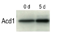
Arabidopsis thaliana wild ecotype Columbia was grown for four weeks under continuous illumination and then transferred to complete darkness for five days. Several leaves were harvested from the plants before they were transferred to darkness (0 d) or after they were kept for five days (5 d). Protein was extracted with the SDS extraction solution containing 50 mM Tris (pH 6.8), 10% (w/v) glycerol, 2% (w/v) SDS and 6% (v/v) 2-mercaptoethanol. Protein extract equivalent to 1 mg leaf material was loaded and separated on 14% SDS-PAGE and blotted 1h to PVDF. Blots were blocked with PBS-T containing 1.5% skim milk for 1h at room temperature (RT) with agitation. Blot was incubated in the primary antibody at a dilution of 1: 10 000 for 1h at RT with agitation. The antibody solution was decanted and the blot was rinsed briefly twice, then washed once for 15 min and 3 times for 5 min in PBS-T at RT with agitation. Blot was incubated in secondary antibody (anti-rabbit IgG horse radish peroxidase conjugated, from GE Healthcare ) diluted to 1:20 000 in for 1h at RT with agitation. The blot was washed as above and developed for 1 min with ECLplus according to the manufacturers instructions. Exposure time was 5 min.
Arabidopsis thaliana wild ecotype Columbia was grown for four weeks under continuous illumination. Several young (1), mature (2) and senescing (3) leaves were harvested from the plants. Protein was extracted with the SDS extraction solution containing 50 mM Tris (pH 6.8), 10% (w/v) glycerol, 2% (w/v) SDS and 6% (v/v) 2-mercaptoethanol. Protein extract equivalent to 1 mg leaf material was loaded and separated on 14% SDS-PAGE and blotted 1h to PVDF. Blots were blocked with PBS-T containing 1.5% skim milk for 1h at room temperature (RT) with agitation. Blot was incubated in the primary antibody at a dilution of 1:30 000 for 1h at RT with agitation as indicated in the figure. The antibody solution was decanted and the blot was rinsed briefly twice, then washed once for 15 min and 3 times for 5 min in PBS-T at RT with agitation. Blot was incubated in the secondary antibody provided by AgriSera (AS09 602) diluted to 1:20 000 in for 1h at RT with agitation. The blot was washed as above and developed for 1 min with ECLplus according to the manufacturers instructions. Exposure time was 5 min.
Courtesy of Kaori Takahashi at Hokkaido University, Japan - Additional Information
-
Additional information: The protein level is moderately induced during dark-induced senescence Additional information (application): This antibody works on total cell extracts and can be used as a senescence marker. Predicted size of Acd1 precursor protein is about 61 kD including the transit peptide, but it must be processed to a smaller size. Using fresh extracts is recommended to decrease possible cross-reaction with Rubisco.
- Background
-
Background: Acd1 (accelerated cell death 1) EC=1.14.12.20 is an enzyme involved in chlorophyll breakdown, pheophorbide a oxygenase. This enzyme seems to induce cell death in Arabidopsis leaves in the dark. Synonymes:PaO, Lls1, lethal leaf-spot 1 homolog, pheide a oxygenase
- Product Citations
-
Selected references: James et al. (2025). A Brassica napus water soluble chlorophyll binding protein (WSCP1) delays chlorophyll degradation and inhibits serine proteases during dark-induced leaf senescence in Arabidopsis thaliana. Planta. 2025 Jul 2;262(2):39. doi: 10.1007/s00425-025-04754-6.
Fukura et al. (2021) Enrichment of chlorophyll catabolic enzymes in grana margins and their cooperation in catabolic reactions. J Plant Physiol. 2021 Nov;266:153535. doi: 10.1016/j.jplph.2021.153535. Epub 2021 Sep 25. PMID: 34607178.
Kim et al. (2013). Mutation of the Arabidopsis NAC016 Transcription Factor Delays Leaf Senescence.'Plant Cell Physiol. Aug 21.
Nagane et al. (2010). Involvement of AtNAP1 in thre reulation of chlorophyll degradation in Arabiopsis thaliana. Planta (4):939-949.
Hirashima et al. (2009). Light-independent cell death induced by accumulation of pheophorbide a in Arabidopsis thaliana. Plant Cell Physiol. (4):719-729. - Reviews:
-
This product doesn't have any reviews.
Related products

AS09 602 | Clonality: Polyclonal | Host: Goat | Reactivity: Rabbit IgG (H&L)
AS06 122 | Clonality: Polyclonal | Host: Rabbit | Reactivity: C.reinhardtii, purple bacteria (CRD1) and plants (CHL27) including A.thaliana, H.vulgare, N. tabacum, P.sativum, P. patens, Synechocystis sp. PCC6803
compartment marker of chloroplast thylakoid and envelope membranes

AS05 067 | Clonality: Polyclonal | Host: Rabbit | Reactivity: Arabidopsis thaliana, Cyanobacteria, Horderum vulgare, Nicotiana tabacum, Oryza sativa, Phalenopsis Sogo Yukidian cultivar V3, Pinus yunnanensis, Pisum sativum, Triticum aestivum