1

Anti-GOGAT | Glutamine oxoglutarate aminotransferase
AS07 242 | Clonality: Polyclonal | Host: Rabbit | Reactivity: A. thaliana, G. max, N. tabacum, O. sativa, Populus sp., S. lycopersicum, S. elongatus, S. oleracea , Z. mays
Benefits of using this antibody
- Product Info
-
Immunogen: KLH-conjugated synthetic peptide well conserved in known GOGAT sequences from different species including Arabidopsis thaliana Fd-GOGAT 1 Q9ZNZ7, At5g04140 and Fd-GOGAT 2 Q9T0P4, At2g41220
Host: Rabbit Clonality: Polyclonal Purity: Serum Format: Lyophilized Quantity: 50 µl Reconstitution: For reconstitution add 50 µl of sterile water Storage: Store lyophilized/reconstituted at -20°C; once reconstituted make aliquots to avoid repeated freeze-thaw cycles. Please remember to spin the tubes briefly prior to opening them to avoid any losses that might occur from material adhering to the cap or sides of the tube. Tested applications: Western blot (WB) Recommended dilution: 1 : 1000 (WB) Expected | apparent MW: 177 | 170-180 kDa depending upon the species
- Reactivity
-
Confirmed reactivity: Arabidopsis thaliana, Miscanthus giganteus, Nicotiana tabacum, Oryza sativa, Phaseolus vulgaris, Populus tremula, Solanum lycopersicum, Spartina sp., Zea mays
Predicted reactivity: Arthrospira platensis PCC 7345, Beta vulgaris, Brachypodium distachyon, Burkholderia glumae, Camellia sinensis, Chloroflexi bacterium, Chlamydonas reinhardtii, Gracilaria tenustipitata, Cyanobacteria, Crocosphaera watsonii WH 8502, Cyanidioschyzon merolae (strain 10D), Galdieria sulphuraria , Glycine max, Helicosporidium sp. ATCC 50920, Hydrogenobaculum sp. HO, Lactococcus lactis subsp. lactis bv. diacetylactis str. TIFN2, Leptospira interrogans serovar Bataviae str. HAI135, Leptolyngbya boryana, Medicago trucutula, Microcystis panniformis, Monoraphidium neglectum, Morus notabilis, Nannochloropsis gaditana, Nicotiana tabacum, Ostreococcus lucimarinus,Physcomitrium patens subsp. patens, Porphyra purpurea, Sutterella wadsworthensis, Sulfurihydrogenibium yellowstonense, Veillonella atypica
Species of your interest not listed? Contact usNot reactive in: No confirmed exceptions from predicted reactivity are currently known - Application Examples
-

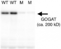
Approximately 40 µg of total protein from Arabidopsis thaliana wild-type (WT) and gls1-103 mutant (M) leaves, the latter of which showed reduced GOGAT levels, was extracted with a protein extraction solution containing 50 mM Imidazole/HCl (pH 7.0 at 4ºC), 20% glycerol, 5mM 6-aminocaproic acid, 1mM EDTA. Protein extracts were then separated on 6% SDS-PAGE and blotted overnight at 4ºC to PVDF using the tank transfer method. Blots were blocked with PBS-T containing 5% skim milk for 1h at room temperature (RT) with agitation. Blot was incubated in the primary antibody at a dilution of 1: 20 000 in PBS-T containing 1% (w/v) BSA for 1h at RT with agitation. The antibody solution was decanted and the blot was rinsed briefly twice, then washed once for 15 min and 3 times for 5 min in PBS-T at RT with agitation. Blot was incubated in secondary antibody (anti-rabbit IgG horse radish peroxidase conjugated) diluted to 1:20 000 in PBS-T containing 1% (w/v) BSA for 1h at RT with agitation. The blot was washed as above and developed for 1 min with chemiluminescent detection reagent, according to the manufacturer's instructions. Exposure time was 5 min.Courtesy Dr. Atsushi Takabayashi, Institute of Low Temperature Science, Hokkaido University, Japan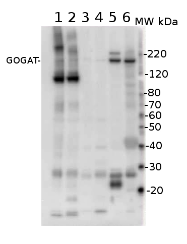
20 µg of total protein cell extract extracted with Protein Extration Buffer, PEB (AS08 300) from leafs of (1) Spartina patens (2) Spartina alterniflora l, (3) Miscanthus giganteus , (4) Zea mays, (5) Phaseolus vulgaris, (6) Arabidopsis thaliana were separated on 4-12% NuPage (Invitrogen) LDS-PAGE and blotted 1h to PVDF. Blots were blocked immediately following transfer in 2% blocking reagent in 20 mM Tris, 137 mM sodium chloride pH 7.6 with 0.1% (v/v) Tween-20 (TBS-T) for 1h at room temperature with agitation. Blots were incubated in the primary antibody at a dilution of 1: 10 000 for 1h at room temperature with agitation. The antibody solution was decanted and the blot was rinsed briefly twice, then washed once for 15 min and 3 times for 5 min in TBS-T at room temperature with agitation. Blots were incubated in secondary antibody (anti-rabbit IgG horse radish peroxidase conjugated) diluted to 1:50 000 in 2% blocking solution for 1h at room temperature with agitation. The blots were washed as above and developed for 5 min with chemiluminescent detection reagent according the manufacturers instructions. Images of the blots were obtained using a CCD imager (FluorSMax, Bio-Rad) and Quantity One software (Bio-Rad). - Additional Information
-
Additional information (application): This antibody can be used as a plastidial marker for leaf material, not roots where levels of GOGAT are too low.
A 40 kDa band present in A. thaliana sample is not competed away during antibody neutralization assay. In this assay free peptide used for antibody production is incubated together with anti-GOGAT antibodies. Due to the MW of this protein we suggest to use a gradient gel for protein separation and a longer transfer time. - Background
-
Background: Glutamine oxoglutarate aminotransferase (abbreviated as GOGAT) is an enzyme involved in synthesis of glutamate from glutamine and alpha-ketoglutarate. GOGAT has two forms in plants: ferredoxin-dependent GOGAT (Fd-GOGAT) and NADH-dependent GOGAT (NADH-GOGAT). 95% of GOGAT found in plants is the Fd-GOGAT type. Fd-GOGAT is encoded by two genes, glu1 and glu2 found on chromosomes 5 and 2 respectively (in Arabidopsis). Fd-GOGAT (both forms) is highly conserved among plants, red algae, and cyanobacteria.
- Product Citations
-
Selected references: Gilad et al. (2025). Nitrogen Assimilation Plays a Role in Balancing the Chloroplastic Glutathione Redox Potential Under High Light Conditions.Plant Cell Environ. 2025 Jan 9.doi: 10.1111/pce.15368.
Rubio Wilhelmi et al. (2024). Salinity-Induced Photorespiration in Populus Vascular Tissues Facilitate Nitrogen Reallocation. Plant Cell Environ. 2024 Oct 1.doi: 10.1111/pce.15180.
Touraine et al. (2019). Iron-sulfur protein NFU2 is required for branched-chain amino acid synthesis in Arabidopsis roots. J Exp Bot. 2019 Mar 27;70(6):1875-1889. doi: 10.1093/jxb/erz050.
Wang et al. (2018). Genetic variations in ARE1 mediate grain yield by modulating nitrogen utilization in rice. Nat Commun. 2018 Feb 21;9(1):735. doi: 10.1038/s41467-017-02781-w.
Nath et al. (2016). A Nitrogen-Fixing Subunit Essential for Accumulating 4Fe-4S-Containing Photosystem I Core Proteins. Plant Physiol. 2016 Dec;172(4):2459-2470. Epub 2016 Oct 26.
Jayawardena et al. (2016). Elevated CO2 plus chronic warming reduces nitrogen uptake and levels or activities of nitrogen -uptake and -assimilatory proteins in tomato roots. Physiol Plant. 2016 Nov 28. doi: 10.1111/ppl.12532. [Epub ahead of print]
Takabayashi et al. (2016) Direct interaction with ACR11 is necessary for post-transcriptional control of GLU1-encoded ferredoxin-dependent glutamate synthase in leaves. Sci Rep. 2016 Jul 14;6:29668. doi: 10.1038/srep29668.
Yang et al. (2016). Rice Ferredoxin-Dependent Glutamate Synthase Regulates Nitrogen-Carbon Metabolomes and Is Genetically Differentiated between japonica and indica Subspecies. Mol Plant. 2016 Sep 24. pii: S1674-2052(16)30195-2. doi: 10.1016/j.molp.2016.09.004.
Moscatelli et al. (2015). Characterisation of detergent-insoluble membranes in pollen tubes of Nicotiana tabacum (L.). Biol Open. 2015 Feb 20. pii: BIO201410249. doi: 10.1242/bio.201410249.
Podgórska et al. (2013). Long-term ammonium nutrition of Arabidopsis increases the extrachloroplastic NAD(P)H/NAD(P)+ ratio and mitochondrial reactive oxygen species level in leaves but does not impair photosynthetic capacity. Plant Cell Environ. April 10. - Protocols
-
Agrisera Western Blot protocol and video tutorials
Protocols to work with plant and algal protein extracts
Agrisera Educational Posters Collection
- Reviews:
-
This product doesn't have any reviews.
Related products

AS09 602 | Clonality: Polyclonal | Host: Goat | Reactivity: Rabbit IgG (H&L)

AS09 607 | Clonality: Polyclonal Host: Goat Reactivity: Rabbit IgG (H&L)
AS05 068 | Clonality: Polyclonal | Host: Rabbit | Reactivity: Lupinus luteus
AS07 242 | Clonality: Polyclonal | Host: Rabbit | Reactivity: A. thaliana, G. max, N. tabacum, O. sativa, Populus sp., S. lycopersicum, S. elongatus, S. oleracea , Z. mays
Benefits of using this antibody