1
Anti-Rhamnogalacturonan-I backbone (clone CCRC-M35)
AS16 3224-1ml | Clonality: monoclonal | Host: Mouse | Reactivity: Arabidopsis thaliana
From the laboratory of Michael G. Hahn, PhD, University of Georgia
- Product Info
-
Immunogen: MeBSA-conjugated Arabidopsis thaliana seed mucilage (Rhamnogalacturonan I), non-covalent complex. Epitope structure for carabohydrate antigen: Rha-(1,4)-GalA-(1,2)-Rha-(1,4)-GalA-(1,2)-Rha-(1,4).
Sub class: IgM Host: Mouse Clonality: Monoclonal Purity: Cell culture supernatant. Format: Liquid Quantity: 1 ml Storage: Antibody can be stored up to 1 month at 4°C, and at -80°C for up to 1 year. Make aliquots to avoid repeated freeze-thaw cycles. Please remember to spin the tubes briefly prior to opening them to avoid any losses that might occur from material adhering to the cap or sides of the tube. Tested applications: ELISA (ELISA), Immunohistochemistry (IHC), Immunofluorescence (IF) Recommended dilution: Undiluted or at 1 : 10 (ELISA), (IHC), (IF) - Reactivity
-
Confirmed reactivity: Arabidopsis thaliana Predicted reactivity: Dicots
Species of your interest not listed? Contact usNot reactive in: No confirmed exceptions from predicted reactivity are currently known - Application Examples
-
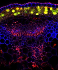
Application example 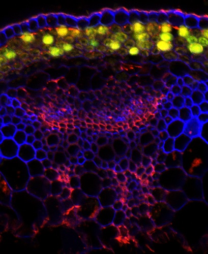
Localization of rhamnogalacturonan-I backbone (red) in Arabidopsis thaliana hypocotyl, Calcufluor White counterstain (blue) and cell wall autofluorescence (yellow).
The 31 days-old hypocotyls were immersed in 150 μL PME fixation buffer (25 mM PIPES, 1 mM MgSO4, 1 mM EGTA) and then subjected to three consecutive cycles of 5 min-long vacuum infiltration (21°C, 68 kPa). Afterwards they were washed three times in PME (21°C, 68 kPa) prior to storage at 4°C in PME. Hypocotyls were encased in 1 cm3 blocks of 5% agar at 65°C, and stored at 4°C to set. Transverse 40 μm thick sections were cut from segments using a VT100S vibrating microtome (Leica) and blocked for at least 1 h in 5% bovine serum albumin in TBST. Blocking solution was discarded and sections were incubated at 4°C for 16 h with 5 μl of the anti-Rhamnogalacturonan-I backbone antibody, followed by 2 washes in 100 μL TBST. Sections were then incubated for 1 h at 21°C in the dark in 10 μl of 2 μg/μl Alexa FluorTM 568 donkey anti-mouse IgG (H+L; 1:36). Sections were again washed twice in 40 μL TBST prior to counter-staining with 0.015% Calcofluor White (Sigma-Aldrich). Sections were again washed twice in 100 μL TBST to remove excess counter-stain and unbound secondary antibody. Immunofluorescence of AlexaFluor 568 was excited with a 561 nm laser, and emitted light filtered at 575–600 nm, while Calcufluor White was subsequently scanned on an independent channel with a 405 nm laser and emission observed at 420–430 nm using laser scanning microscope Zeiss LSM780 point-scan system at 1024 × 1024 pixels (pixel size, 0.6–0.83 μm) with a 10X objective.
Courtesy Dr. Urs Fisher, Umeå Plant Science Centre, Sweden - Additional Information
-
Additional information: Exact working dilution needs to be determined by end user Additional information (application): CCRC-M35 binds to the backbone of rhamnogalacturonan I and requires at least two unbranched disaccharide repeats for binding, CCRC-M35 does not bind to branched sections of the backbone and is not sensitive to the identity of the sugar at the non-reducing terminus - Background
-
Background: Rhamnogalacturonans (RGs) are pectic polysaccharides found in the cell wall, which contain a repeating dissacharides backbone: α-D-GalpA-(1,2)-α-L-Rhap-(1)
The antibody was raised against seed mucilage/MeBSA complex and binds to Rha-(1,4)-GalA-(1,2)-Rha-(1,4)-GalA-(1,2)-Rha-(1,4) pectic backbone. - Product Citations
-
Selected references: Pattathil et al. (2012). Immunological approaches to plant cell wall and biomass characterization: Glycome Profiling. Methods Mol Biol. 2012;908:61-72.doi: 0.1007/978-1-61779-956-3_6.
Patathil et al. (2010). A comprehensive toolkit of plant cell wall glycan-directed monoclonal antibodies. Plant Physiol. 2010 Jun;153(2):514-25.doi: 10.1104/pp.109.151985.
- Reviews:
-
This product doesn't have any reviews.