3

Anti-PIP1;3 | Aquaporin, plasma membrane intrinsic protein 1-3
AS22 4811 | Clonality: Polyclonal | Host: Rabbit | Reactivity: Arabidopsis thaliana, Cardamine hirsuta,Oryza sativa, Zea mays
- Product Info
-
Immunogen: KLH-conjugated peptide derived from Oryza sativa PIP1;3 protein sequence, UniProt: Q9SXF8 Host: Rabbit Clonality: Polyclonal Purity: Antigen affinity purified serum, in PBS pH 7.4 Format: Lyophilized Quantity: 50 µg Reconstitution: For reconstitution, add 50 µl, of sterile or deionized water. Storage: Store lyophilized/reconstituted at -20°C; once reconstituted make aliquots to avoid repeated freeze-thaw cycles. Please, remember to spin tubes briefly prior to opening them to avoid any losses that might occur from lyophilized material adhering to the cap or sides of the tubes. Tested applications: Immunofluorescence (IF), Western blot (WB) Recommended dilution: 1: 500 (IF), 1 : 1000 (WB) Expected | apparent MW: 26-29 kDa (Oryza sativa) - Reactivity
-
Confirmed reactivity: Arabidopsis thaliana, Cardamine hirsuta, Oryza sativa, Zea mays Predicted reactivity: Alium sativum, Glycine max, Hordeum vulgare, Miscanthus floridulus, Setaria italica, Triticum aestivum, Triticum urartu
Species of your interest not listed? Contact usNot reactive in: Nicotiana benthamiana, Physcomitrium patens, Solanum lycopersicum - Application Examples
-
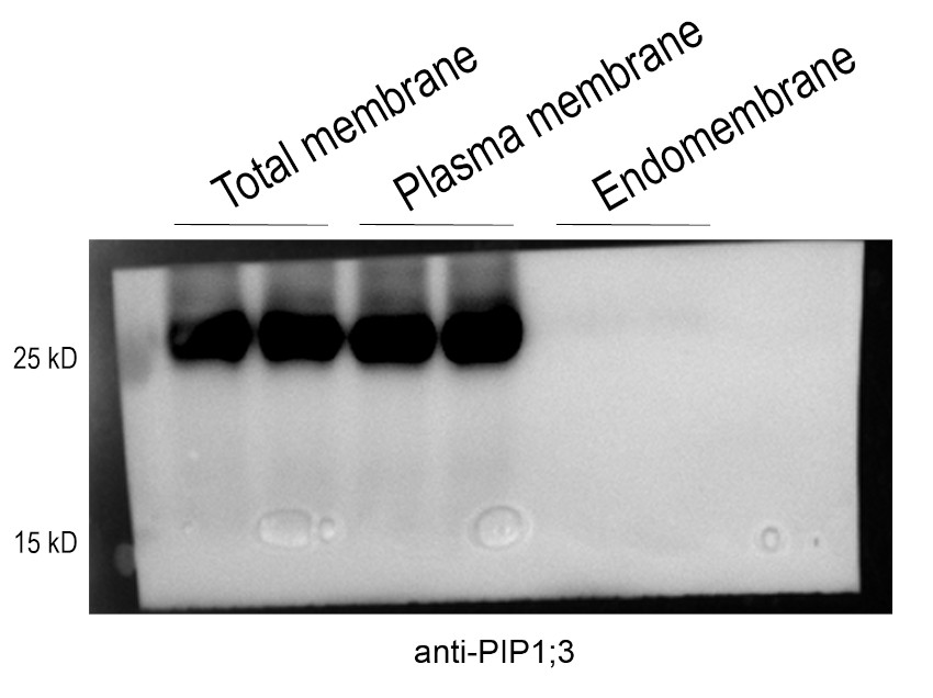
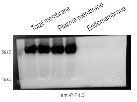
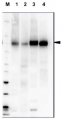
Samples:
Membrane proteins extracted from 3-week-old Oryza sativa plants, specifically the proteins collected and concentrated from the PEG fractions (containing plasma membrane protein) and DEX fractions (cAS22 ontaining endomembrane protein) during the microsomal protein fractionation process.
About 10 µg/well of membrane proteins (extracted from 3-week-old rice plants) were denatured in the corresponding buffer mixed with 6×Protein Loading Buffer at 37 °C for 10 min. Samples were separated at room temperature on 10% SDS-PAGE and blotted for 1 h to nitrocellulose (pore size of 0.45 um) using wet transfer in the cold. The blot was blocked with 5% milk for 1 h at room temperature with agitation. The blot was incubated in the primary antibody at a dilution of 1:5000 for 1 h at room temperature with agitation in TBS-T. The antibody solution was decanted and the blot was rinsed briefly three times, then washed 3 times for 5 min each in TBS-T at room temperature with agitation. The blot was incubated in the matching secondary antibody (anti-rabbit IgG horse radish peroxidase conjugated) diluted to 1:10000 for 1 h at room temperature with agitation. The blot was washed as above and developed with the chemiluminescent detection reagent: Tanon High-sig ECL Western Blotting Substrate. The exposure time was 10 seconds.Courtesy of Dr. Shuoxun Wang, Institute of Genetics and Developmental Biology, Chinese Academy of Sciences, Beijing, China
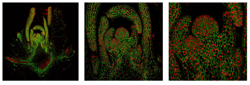
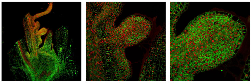
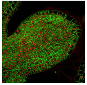
Material: shoots of Arabidopsis thaliana (first set of images) and Cardamine hirsuta (second set of images)
The following protocol was applied:
Fixation: FAA: 4% formaldehyde, 50% ethanol, 5 % acetic acid (A. thaliana)
PFA: 4% formaldehyde in 50 mM phosphate buffer pH 7.5 (C. hirsuta)
Embedding and sectioning: Steedman wax/10 μm sections
Hydrophilization: no
Cell wall digestion: no
Membrane permeabilization: DMSO-IGEPAL
Antigen retrieval: no
Blocking buffer: TSA blocking reagent (Perkin Elmer)
Washing buffer: PBS with 0.1% gelatine
Primary antibody: 1: 500 incubation o/n at 4°C
Secondary antibody: GAR-alexa488 (ThermoFisher) 1:250, 2h/RT
Co-staining of the nucleus (DAPI): yes
Cell wall staining: no
Imaging: Leica Stellaris 5 confocal microscope
Note: fixation performed with: PFA or FAA was giving comparable results.
Dr. Ton Timmers, Max Planck Institute for Plant Breeding Reseach, Cologne, Germany - Background
-
Background: PIP1s - is a plasma membrane aquaporine which facilitates trasport of water across cell membrane. - Protocols
-
Agrisera Western Blot protocol and video tutorials
Protocols to work with plant and algal protein extracts
Agrisera Educational Poster Collection - Reviews:
-
This product doesn't have any reviews.
Related products

AS09 602 | Clonality: Polyclonal | Host: Goat | Reactivity: Rabbit IgG (H&L)
AS07 260 | Clonality: Polyclonal | Host: Rabbit | Reactivity: [global antibody] for di- and monocots, conifers, ferns, mosses, green algae | Cellular [compartment marker] for plasma membrane
Antibody to phosphorylated H+ATPase
AS21 4697 | Clonality: Monoclonal | Host: Mouse | Reactivity: Zea mays
AS12 2110 | Clonality: Polyclonal | Host: Rabbit | Reactivity: L. sativa, P. sativum, S. lycopersicum, Z. mays