1
Anti-COP1 | E3 ubiquitin-protein ligase COP1
AS20 4399 | Clonality: Polyclonal | Host: Rabbit | Reactivity: Arabidopsis thaliana
- Product Info
-
Immunogen: His-tagged recombinant part of COP1 protein from Arabidopsis thaliana, overexpressed in E.coli, UniProt: P43254, TAIR: AT2G32950 Host: Rabbit Clonality: Polyclonal Purity: Antigen affinity purified serum, in PBS pH 7.4 Format: Lyophilized Quantity: 50 µg Reconstitution: For reconstitution add 50 µl, of sterile water. Storage: Store lyophilized/reconstituted at -20°C; once reconstituted make aliquots to avoid repeated freeze-thaw cycles. Please, remember to spin tubes briefly prior to opening them to avoid any losses that might occur from lyophilized material adhering to the cap or sides of the tubes. Tested applications: Western blot (WB) Recommended dilution: 1 : 1000 (WB) Expected | apparent MW: 75 kDa - Reactivity
-
Confirmed reactivity: Arabidopsis thaliana Predicted reactivity: Brassica napus, Brassica rapa, Camelina sativa, Capsella rubella, Eutrema salsugineum, Hirschfeldia incana, Raphanus sativus, Solanum lycopersicum
Species of your interest not listed? Contact us
Not reactive in: No confirmed exceptions from predicted reactivity are currently known - Application Examples
-
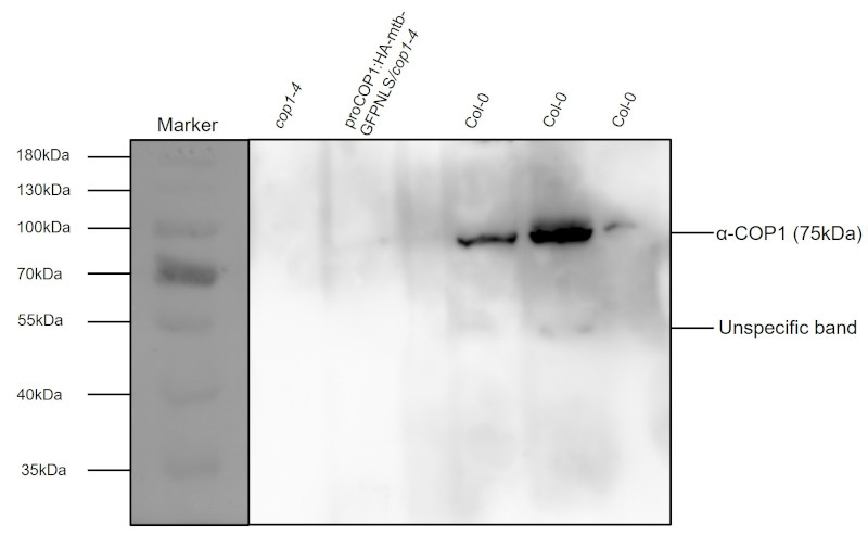
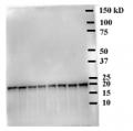
Protein loading: 50 µg total protein per well.
Sample source: Arabidopsis thaliana, 4-day-old dark-grown seedlings. Negative control was cop1-4 mutant.
Samples denatured at 95°C for 10 minutes (in standard SDS sample buffer) and conducted to SDS-PAGE in 10% gel followed by Semi-dry transfer (Bio-Rad fast blotting system) using PVDF membrane (0.45 µm pore size) for 30 minutes at RT. Blocking was done using 1× Roti-Block (Carl Roth) for 1 h/RT with agitation. Primary antibody was incubated at dilution 1: 1000 in 2 % nonfat milk in 1xPBS ON/4°C with agitation. The blot was washed in the following way: 1 × 15 min wash + 3 × 5 min washes in TBS-T at RT with agitation followed by incubation in the secondary antibody Anti-rabbit IgG (HRP-conjugated) at 1: 50 000 for 1h/RT with agitation. Detection was performed using mid-femtogram detection range chemiluminescent detection reagent. Exposure time was few seconds.Courtesy of prof. Ute Hoecker group, University of Cologne, Germany
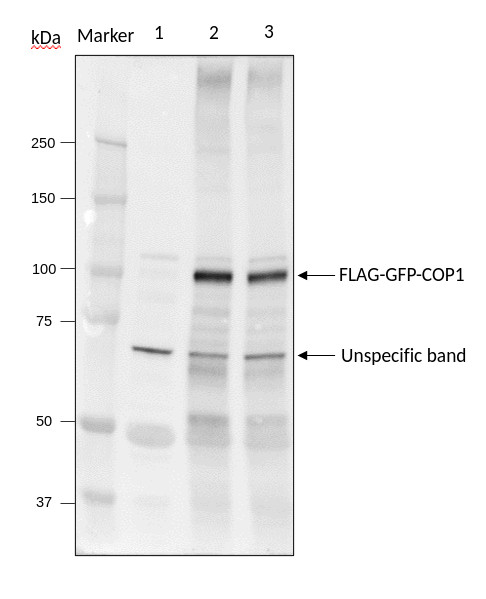
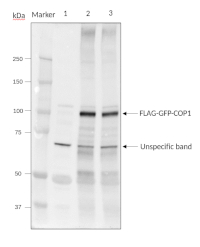
Samples:
Marker: Precision Plus Protein Dual Color Standards (Biorad, #1610394)
1 - Arabidopsis thaliana cop1-4 mutant
2 - Arabidopsis thaliana 35S-FLAG-GFP-COP1/cop1-4 transgenic line #1
3 - Arabidopsis thaliana 35S-FLAG-GFP-COP1/cop1-4 transgenic line #2 Total protein extracted freshly from 15 seedlings (7-day-old grown under short day condition with 8h light/16h dark) with 2x Laemmli sample buffer (Biorad, 120 mM Tris-HCl pH 6.8, 4% SDS, 20% glycerol, 0.02% bromophenol blue, 200 Mm dithiothreitol), and then denatured at 95°C for 10 min. Proteins were separated on pre-cast SDS-polyacylamide gels with a 7.5% acrylamide and blotted 7 min to nitrocellulose membranes (Biorad Trans-Blot Turbo RTA Nitrocellulose Transfer Kit), using semi-dry transfer. Blot was blocked with EveryBlot Blocking Buffer (Biorad) for 10 min at RT with agitation. Bolt was incubated in the anti-COP1 antibody at a dilution of 1:1000 for 4 °C/ON with agitation. The primary antibody solution was decanted, and the membrane was first washed briefly once (5 s) and then for 15 min, followed by 3 additional washings of 5 min in 1x TBS-T (without blocking agent) with agitation. Blot was incubated in Agrisera matching secondary antibody (Goat anti-Rabbit IgG, HRP conjugated, AS09 602) diluted to 1:25000 for 1h at RT with agitation). The blot was washed as above and developed for 2~3 min with chemiluminescent detection (AgriseraSuperBright, AS16 ECL-S-10), according to the manufacture’s instructions. The Exposure time was 20 seconds.
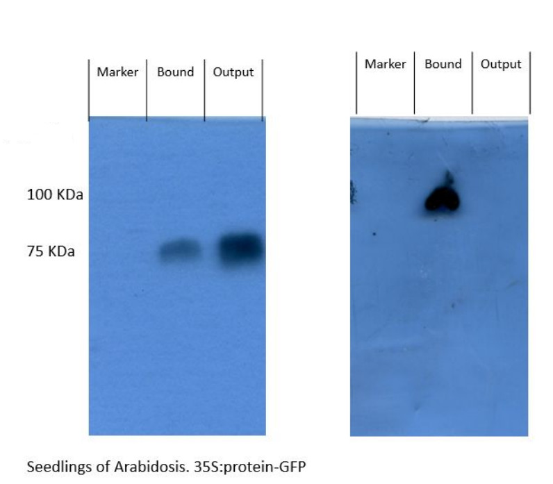
Left blot: Anti-COP1 antibody (AS20 4399). Right blot anti-GFP antibody (Agrisera).
The protocol described below enables the isolation of protein complexes through Co-IP and their subsequent detection by Western blot. The INPUT, OUTPUT, and BOUND fractions allow evaluation of immunoprecipitation efficiency and detection of proteins of interest.Procedure:
Materials and General Conditions
● Previously ground plant tissue
● Lysis buffer
● Cold dilution buffer
● Immunoprecipitation beads
● Glycine 0.2 M pH 2.5
● Tris Base (0.6 M or 1 M, pH 10.8)
● Sample Buffer (SB)
● Blocking solution (skim milk in TBS-T)
● Primary antibody
● Secondary antibody (anti-rabbit)
● TBS-T buffer
● Conditions: Perform the protocol on ice or at 4°CProcedure
1. Cell Lysis
420 µl of lysis buffer were added to the previously ground plant tissue. The mixture was incubated on ice for 30 minutes with gentle mixing by inversion (or in a cold shaker). The sample was centrifuged at 12,500 RPM and 5°C for 10 minutes.
2. Bead Equilibration
Beads were gently resuspended using a pipette (DO NOT VORTEX). 25 µl per sample were transferred into 1.5 ml tubes. 500 µl of cold dilution buffer were added and samples were equilibrated for approximately 30 minutes.
3. Protein Extract Preparation
Approximately 350 µl of supernatant were transferred to a clean tube. 1 ml of dilution buffer was added to the remaining supernatant, followed by centrifugation at 12 500 RPM and 5°C for 10 minutes.
4. Immunoprecipitation
The resulting supernatant (~1 ml) was transferred into a tube containing equilibrated beads (after carefully removing the buffer without losing the beads). 200 µl were separated as the INPUT fraction into a new tube. Samples were incubated for 1 hour at 4°C with head-to-tail rotation.
5. Separation and Washing
Samples were placed on a magnet to separate beads.
● The supernatant was collected as the OUTPUT fraction, mixed with 1 volume of SB,and stored.
● The bead pellet was washed with 500 µl of cold dilution buffer for 2 minutes. This washing step was repeated three times.A quick spin was performed, followed by magnetic separation and removal of the supernatant.
6. Elution of Complexes
Protein complexes were eluted using 15 µl of glycine 0.2 M pH 2.5, with gentle shaking for 30 seconds.Elution was neutralized with 3 µl of Tris Base (0.6 M or 1 M, pH 10.8). 20 µl of SB were added, resulting in the BOUND fraction.
7. Sample Denaturation
Samples were heated at 100°C for 10 minutes and stored at −20°C until gel loading. Following gel electrophoresis and protein transfer, the membrane was blocked with blocking solution (5% skim milk in TBS-T) for 1 hour and 30 minutes at room temperature with gentle agitation, followed by incubation with primary antibody performed overnight at 4°C (2.5 µl in 10 ml of 1% milk in TBS-T) with gentle agitation. Membrane was washed with TBS-T for 5 minutes, three times, followed by a secondary antibody incubation with anti-rabbit secondary antibody (2.5 µl in 10 ml of 2.5%milk in TBS-T) for 1 h/RT. Signal detection was performed using radiographic film.Courtesy of Ana Medina Fraga, Lic en Biotecnología PhD student Ballaré Lab CONICET-IFEVA-UBA-UNSAM, Argentina
- Background
-
Background: COP1 (E3 ubiquitin-protein ligase COP1) is onvolved in proteasome-mediated ubiquitin-dependent protein catabolic process and protein ubiquitination. The protein contains a ring finger zinc-binding motif, a coiled-coil domain, and several WD-40 repeats, similar to G-beta proteins. Acts as a repressor of photomorphogenesis and as an activator of etiolation in darkness. Represses photomorphogenesis in darkness by mediating ubiquitination and subsequent proteasomal degradation of light-induced transcription factors such as HY5, HYH and LAF1. Localized in the nucleus in darkness but is gradually relocated to the cytoplasm upon illumination.
Alternative names: Constitutive photomorphogenesis protein 1, RING-type E3 ubiquitin transferase COP1 - Protocols
-
Agrisera Western Blot protocol and video tutorials
Protocols to work with plant and algal protein extracts
Agrisera Educational Poster Collection - Reviews:
-
This product doesn't have any reviews.
Related products

AS09 602 | Clonality: Polyclonal | Host: Goat | Reactivity: Rabbit IgG (H&L)
AS12 1867 | Clonality: Polyclonal | Host: Rabbit | Reactivity: Arabidopsis thaliana