Visar bild 1 av
1
Anti-H3S10p | Histone H3 (p Ser10) (affinity purified)
Product no: AS16 3635
AS16 3635 | Clonality: Polyclonal | Host: Rabbit | Reactivity:Human
427
€
Customer reviews
Delivery:
3-6 business days
- Product Info
-
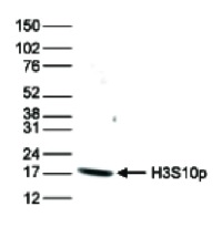
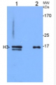
Immunogen: KLH-conjugated synthetic peptide Host: Rabbit Clonality: Polyclonal Purity: Immunogen affinity purified serum. Format: Liquid Quantity: 50 µg Storage: Store lyophilized/reconstituted at -20°C; once reconstituted make aliquots to avoid repeated freeze-thaw cycles. Please remember to spin the tubes briefly prior to opening them to avoid any losses that might occur from material adhering to the cap or sides of the tube. Tested applications: Chromatin immunoprecipitation (ChIP), Dot Blot (Dot), ELISA (ELISA), Immunofluorescence (IF), Western blot (WB) Recommended dilution: 2 µg/million cells (ChIP), 1 : 20 000 (Dot), 1 : 2000 (IF), 1 : 100 (WB) Expected | apparent MW: 15 | 17 kDa
- Reactivity
-
Confirmed reactivity: Human Predicted reactivity: Chicken, Drosophila melanogaster, Mouse, Plant, Rat, Xenopus sp. Not reactive in: No confirmed exceptions from predicted reactivity are currently known - Application Examples
-
application example 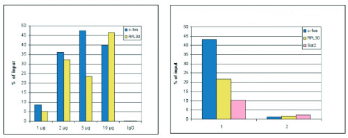
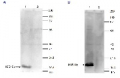
ChIP: results obtained with anti-H3S10p polyclonal antibodies.
Left panel: ChIP assays were performed using human HeLa cells treated with colcemid to block the cells in metaphase, anti-H3S10p antibodies and optimized PCR primer sets for qPCR. ChIP was performed on sheared chromatin from 10 000 cells using the SX-8G IP-Star automated system. A titration of the antibody consisting of 1, 2, 5, and 10 μ g per ChIP experiment was analysed. IgG (5 μ g/IP) was used as negative IP control. QPCR was performed with primers for the promoter of the active genes c-fos and RPL30. Figure shows the recovery, expressed as a % of input (the relative amount of immunoprecipitated DNA compared to input DNA after qPCR analysis).
Right panel: ChIP was performed as described above using 2 μ g of H3S10p antibody and sheared chromatin from 10 000 HeLa cells treated with colcemid (sample 1) or from 10 000 untreated cells (sample 2). QPCR was performed with primers for the promoter of the active genes c-fos and RPL30, and for the Sat2 satellite repeat region.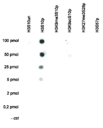
Dot Blot: analysis was performed to test the cross reactivity of this antibody with peptides containing other modifications of histone H3 or the unmodified H3S10 sequence. One hundred to 0.2 pmol of the peptide containing the respective histone modification were spotted on a membrane. The antibody was used at a dilution of 1:20 000. Image shows a high specificity of the antibody for the modification of interest. The antibody does not recognize the H3S10p modification if the neighboring K9 is acetylated or trimethylated.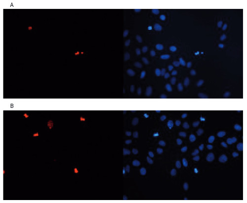
Immunofluorescence: using anti-H3S10p antibodies. Human osteosarcoma (U2OS) cells were stained with this antibody and with DAPI. Cells were fixed with 3.7% formaldehyde in PBS for 20’ at RT, followed by a 20’ permeabilization with 0.5% Triton X-100 in PBS and blocked PBS/TX-100 containing 5% normal goat serum Figure 5A: cells were immunofluorescently labeled with the H3S10p antibody (left), diluted 1:2 000 in blocking buffer followed by an anti-rabbit antibody conjugated to Alexa568 or with DAPI (right), which specifically labels DNA. Figure 5B: staining of the cells with the H3S10p antibody after incubation of the antibody with 2 μM blocking peptide containing the unmodified H3S10 sequence, the phosphorylated H3S10 and the phosphorylated H3T11, respectively.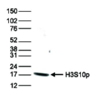
Western blot: was done using anti-H3S10p antibodies and HeLa cells were treated with colcemid, which blocks the cell cycle in metaphase and 15 μ g of histone extracts of the cells were analysed by Western blot this antibody diluted 1:1 000 in TBS-Tween containing 5% skimmed milk. The position of the protein of interest is indicated on the right; the marker (in kDa) is shown on the left. - Additional Information
-
Additional information: This antibody preparation is provided in 20 mM Potassium Phosphate pH 7,2, 150 mM NaCl, 0,05% sodium azide and 0,05% ProClin300 - Background
-
Background: Histones are the main constituents of the protein part of chromosomes of eukaryotic cells. They are rich in the amino acids arginine and lysine and have been greatly conserved during evolution. Histones pack the DNA into tight masses of chromatin. Two core histones of each class H2A, H2B, H3 and H4 assemble and are wrapped by 146 base pairs of DNA to form one octameric nucleosome. Phosphorylation of H3S10 is associated with mitosis. - Reviews:
-
This product doesn't have any reviews.
Related products
AS16 3636 | Clonality: Polyclonal | Host: Rabbit | Reactivity:Human
427 €

AS09 602 | Clonality: Polyclonal | Host: Goat | Reactivity: Rabbit IgG (H&L)
219 €
AS10 710 | Clonality: Polyclonal | Host: Rabbit | Reactivity: A. thaliana, B. cinerea, C.annuum, C. reinhardtii, G. hirsutum, H. vulgare, S. lycopersicum, V. faba, P. tricornutum, P. patens, S. europaea, Z. mays | cellular [compartment marker] of nucleoplasm
329 €