1
Anti-PDF1 | Plant defensin 1.1
AS16 3973 | Clonality: Polyclonal | Host: Rabbit | Reactivity: Solanum lycopersicum
- Data sheet
- Product Info
-
Immunogen: KLH-conjugated synthetic peptide derived from Arabidopsis thaliana PDF1.1 UniProt: P30224-1, TAIR: At1g75830
The peptide sequence, it is perfectly conserved in following Arabidopsis thaliana isoforms: PDF1.2c, PDF1.2b, PDF1.2A, PDF1.3. PDF2 isoform sequence did not come in the blast.
Host: Rabbit Clonality: Polyclonal Purity: Affinity purified serum in PBS pH 7.4 Format: Lyophilized Quantity: 50 µg Reconstitution: For reconstitution add 50 µl, of sterile water. Storage: Store lyophilized/reconstituted at -20°C; once reconstituted make aliquots to avoid repeated freeze-thaw cycles. Please, remember to spin tubes briefly prior to opening them to avoid any losses that might occur from lyophilized material adhering to the cap or sides of the tubes.
Tested applications: Western blot (WB) Recommended dilution: 1 : 1000 (WB)
Expected | apparent MW: 8.7 kDa - Reactivity
-
Confirmed reactivity: Arabidopsis thaliana, Solanim lycopersicum
Predicted reactivity: Arabidopsis thaliana, Brassica rapa, Camelina sativa, Eutrema salsugineum, Capsella rubella, Nicotiana tabacum, Sorghum sp.
Species of your interest not listed? Contact usNot reactive in: Nicotiana benthamiana, Solanum tuberosum - Application Examples
-
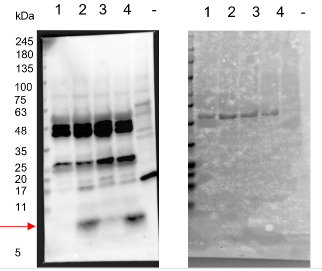
Samples:
Marker used is Prestained Protein SHARPMASS™ VI Protein MW marker (5-245 kDa) from Euroclone company.
sample 1:, 40 µg of Arabidopsis thaliana total protein from wildtype leaves 0 hour post infection with Botrytis cinerea (negative control);
sample 2: 40 µg of Arabidopsis thaliana total protein from mutant leaves (with induced expression of PDF1 mRNA) 0 hour post infection with Botrytis cinerea (positive control);
sample 3: 40 µg of Arabidopsis thaliana total protein from wild type leaves 24 hours post infection with Botrytis cinerea (positive control, PDF1 is expected to be induced by the pathogen used in the experiment);
sample 4: 40 µg of Arabidopsis thaliana total protein from mutant leaves (with induced expression of PDF1 mRNA) leaves 24 hours post infection with Botrytis cinerea (positive control);
samples "-", 10 µg of Arabidopsis thaliana total protein wild type seedlings grown in dark condition (Negative control)
Up to 40 µg/well of total protein extracted freshly from 6-week-old leaves of Arabidopsis thaliana with extraction buffer (125 mm Tris, pH 6.8, 4% [w/v] SDS, 20% [v/v] glycerol, 0.02% [w/v] bromophenol blue, 10% [v/v] β-mercaptoethanol) and denatured 95°C for 7 minutes were separated on a 4–20% Mini-PROTEAN® TGX™ Precast Protein Gels (biorad) SDS-PAGE and blotted 1h to PVDF (pore size of 0.2 µm), using Trans-Blot Turbo Transfer System (Bio-Rad). Blot was blocked with 5% milk for 1h/RT with agitation. Blot was incubated in the primary antibody at a dilution of 1: 1 000 for overnight (~16hours) with agitation in PBS-T with agitation at +4 °C. The antibody solution was decanted and the blot was rinsed briefly twice, then washed once for 15 min and 3 times for 5 min in PBS-T at RT with agitation. Blot was incubated in Agrisera matching secondary antibody (anti-rabbit IgG horse radish peroxidase conjugated) diluted to 1:10 000 for 2h/RT with agitation. The blot was washed as above and developed for 3 min with Agrisera ECL SuperBright with ChemiDoc Imaging Systems (Bio-Rad). Exposure time was 10 seconds.
Left membrane: Western blot detection. Right membrane: staining.
Courtesy Dr. Ricardo Lorrani, University of Rome Sapienza, Italy - Background
-
Background: PDF1 (Plant defensin 1) provides broad-spectrum resistance to pathogens and possesses antifungal activity. Alternative names: Anther-specific protein S18 homolog,Cysteine-rich antifungal protein 1, AFP1,Low-molecular-weight cysteine-rich protein 67, Protein LCR67. - Product Citations
-
Selected references: Nikoloudakis et al. (2020). Structural Diversity and Highly Specific Host-Pathogen Transcriptional Regulation of Defensin Genes Is Revealed in Tomato. Int J Mol Sci. 2020 Dec 9;21(24):9380. doi: 10.3390/ijms21249380. PMID: 33317090; PMCID: PMC7764197. - Protocols
-
Agrisera Western Blot protocol and video tutorials
Protocols to work with plant and algal protein extracts
Agrisera Educational Posters Collection
- Reviews:
-
This product doesn't have any reviews.
Related products

AS09 602 | Clonality: Polyclonal | Host: Goat | Reactivity: Rabbit IgG (H&L)
AS10 687 | Clonality: Polyclonal | Host: Rabbit | Reactivity: A. thaliana, H. vulgare, N. bentamiana, S. oleracea, S. lycopersicum, T. aestivum, Z. mays, V. vinifera
AS16 4107 | Clonality: Polyclonal | Host: Rabbit | Reactivity: Arabidopsis thaliana