1
Anti-PGRL1 | PGR5-like protein 1A (chloroplastic)
AS19 4311 | Clonality: Polyclonal | Host: Rabbit | Reactivity: Arabidopsis thaliana; Picea abies, Pinus sylvestris
- Product Info
-
Immunogen: KLH-conjugated peptide derived from Arabidopsis thaliana PGRL1A UniProt: Q8H112, TAIR: At4g22890
Peptide used to elicit this antibody is conserved in both isoforms PGRL1A and 1B of Zea maysHost: Rabbit
Clonality: Polyclonal Purity: Immunogen affinity purified serum in PBS pH 7.4. Format: Lyophilized Quantity: 50 µg Reconstitution: For reconstitution add 50 µl of sterile water Storage: Store lyophilized/reconstituted at -20°C; once reconstituted make aliquots to avoid repeated freeze-thaw cycles. Please remember to spin the tubes briefly prior to opening them to avoid any losses that might occur from material adhering to the cap or sides of the tube. Tested applications: Western blot (WB) Recommended dilution: 1 : 1000 (WB) Expected | apparent MW: 29 | 29 kDa (Arabidopsis thaliana, Spinacia oleracea) - Reactivity
-
Confirmed reactivity: Arabidopsis thaliana, Picea abies, Pinus sylvestris, Setaria viridis Predicted reactivity: Dichanthelium oligosanthes, Glycine soja, Gossypium arboreum, Klebsormidium nitens, Nelumbo nucifera, Noccaea caerulescens, Physcomitrium patens, Prunus dulcis, Prunus yedoensis, Rhizophora mucronatamm, Solanum chacoense, Trifolium medium, Zea mays, Vitis vinifera
Species of your interest not listed? Contact usNot reactive in: Chlorella sp., diatoms - Application Examples
-
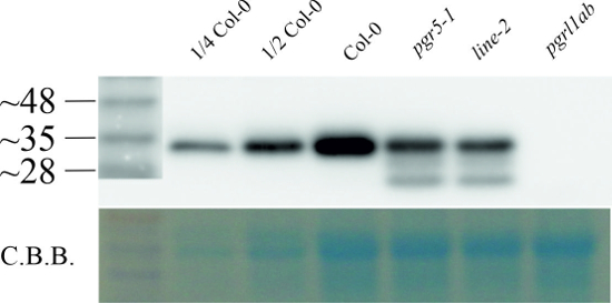
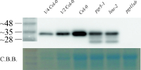
Whole leaf protein (corresponding to 3 mg fresh weight) from the Arabidopsis thaliana lines Columbia-0 (Col-0), PGR5 knock out (pgr5-1), line-2 (line-2) and PGRL1A+B knock out (pgrl1ab) was extracted and denatured with 2x Tricine sample buffer (100mM Tris/HCl, 24% (w/v) glycerol, 8% SDS and 15mM DTT) by heating for 5 min at 70°C. The samples were separated on a 10% Tris Tricine gel and blotted overnight on PVDF-membrane using capillary blotting. The membrane was blocked with 5% milk in Tris-Buffered saline and 0.1% Tween (TBS-T (50mM Tris, 150mM NaCl, 0.1% Tween, pH 7.5)) for 1h at room temperature with agitation and subsequently incubated in the primary antibody at a dilution of 1: 5000 overnight at 4°C with agitation in 1% milk in TBS-T. The antibody solution was decanted and the blot was rinsed briefly, then washed 3 times for 10 min in TBS-T at RT with agitation. The membrane was incubated in secondary antibody (anti-rabbit IgG horse radish peroxidase conjugated, from Agrisera, AS09 602) diluted to 1:10 000 in 1% milk in TBS-T for 1h at RT with agitation. The blot was washed 3 times for 10 min in TBS-T, once for 5 min in TBS (50mM Tris, 150mM NaCl, pH 7.5) and developed for 5 min with chemiluminescent detection reagent according to manufacture's recommendations. Exposure time was 5 seconds.
Courtesy of Jan-Ferdinand Penzler from Ludwig-Maximilians-University Munich (LMU), Dario Leister Lab, Germany
Experimental conditions for detection in extracts of Picea abies and Pinus sylvestris:25 µg/well of total protein extracted from needles of Picea abies and Pinus sylvestris. Exact buffer components were: 10% trichloroacetic acid (TCA)/ acetone buffer. Then followed by a methanol buffer (80% methanol with 0.1 M ammonium acetate) wash and an 80% acetone wash step. Subsequently, total of 1.8 ml mixture of 1:1 phenol (pH 8.0)/SDS buffer (30% sucrose, 2% SDS, 0.1M Tris-HCl, pH 8.0, 5% 2-mercaptoethanol) were added to the tube to extract proteins from dry pellets. After centrifuging at 16,000 g, 4 °C for 3 min, 0.4 ml phenol phase extraction were mixed with 1.6 ml methanol buffer and then centrifuged at 16,000 g, 4 °C for 3 min. The pellets were washed twice with methanol and 80% acetone. Final protein pellets were dissolved in 0.2 ml Laemmli sample buffer, and denatured at 100 °C for 10 min. Samples were separated in the cold on 12 % SDS-PAGE and blotted for 1 h to PVDF membrane, using: wet transfer in the cold. Blot was blocked with 5 % milk for: 1h/RT with agitation. Blot was incubated in the primary antibody at a dilution of 1: 1000 for ON/4°C with agitation. The antibody solution was decanted and the blot was rinsed briefly twice, then washed once for 15 min and 3 times for 5 min in TBS-T at RT with agitation. Blot was incubated in matching secondary antibody (anti-rabbit IgG horse radish peroxidase conjugated) diluted to 1: 10 000 in for 1h/RT with agitation. The blot was washed as above and developed with a following chemiluminescent detection reagent:. Exposure time was 1-5 minutes. Yang et al. 2020.
- Additional Information
-
Additional information (application): This product can be sold containing ProClin if requested - Background
-
Background: PGRL1 is a transmembrane protein present in thylakoids of higher plants and algae. Arabidopsis plants lacking PGRL1 show perturbation of cyclic electron transport (CEF) around photosystem I (PSI), similar to PGR5-deficient plants. PGRL1 has been shown to interact physically with PGR5 and associate with PSI (DalCorso et al., 2008). In Chlamydomonas reinhardtii, PGRL1 is part of a protein supercomplex, composed of PSI with its own light-harvesting complex (LHCI), the photosystem II light-harvesting complex (LHCII), the cytochrome b6/f complex (Cyt b6f), ferredoxin (Fd)-NADPH oxidoreductase (FNR), responsible of the energy balance of the two photosystems and of the switch between thylakoid linear and cyclic electron transport (Iwai et al., 2010).
Synonymes: Ferredoxin-plastoquinone reductase 1 - Product Citations
-
Selected references: McKinnon et al. (2020). Membrane Chaperoning of a Thylakoid Protease Whose Structural Stability Is Modified by the Protonmotive Force. Plant Cell DOI: 10.1105/tpc.19.00797 - Protocols
-
Agrisera Western Blot protocol and video tutorials
Protocols to work with plant and algal protein extracts
Agrisera Educational Poster Collection - Reviews:
-
This product doesn't have any reviews.
Related products

AS09 602 | Clonality: Polyclonal | Host: Goat | Reactivity: Rabbit IgG (H&L)