1
Anti-PsaE | PSI-E subunit of photosystem I (cyanobacterial)
AS19 4355 | Clonality: Polyclonal | Host: Rabbit | Reactivity: Synechocystis sp. PCC 6803, Thermosynechococcus elongatus
- Product Info
-
Immunogen: Recombinant, full length PsaE of Thermosynechococcus elongatus, overexpressed in E.coli, UniProt: P0A423 Host: Rabbit Clonality: Polyclonal Purity: Serum Format: Lyophilized Quantity: 50 µl Reconstitution: For reconstitution add 50 µl of sterile water Storage: Store lyophilized/reconstituted at -20°C; once reconstituted make aliquots to avoid repeated freeze-thaw cycles. Please remember to spin the tubes briefly prior to opening them to avoid any losses that might occur from material adhering to the cap or sides of the tube. Tested applications: Western blot (WB) Recommended dilution: 1 : 5000 - 1: 10 000 (WB) Expected | apparent MW: 8 kDa
- Reactivity
-
Confirmed reactivity: Synechocystis sp. PCC 6803, Thermosynechococcus elongatus Predicted reactivity: Cyanidioschyzon merolae (strain 10D), Synechococcus elongatus PCC 6301, Synechococcus sp. PCC 7002, Trichodesmium erythraeum, Trichormus variabilis - Application Examples
-
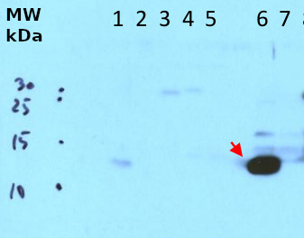
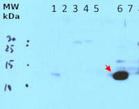
Soluble (8 μg protein) and membrane proteins (corresponding to 1 μg of chlorophyll a) from Synechocystis sp. PCC 6803
1 – WT (soluble)
2 - DPsaE – PsaE deletion (soluble)
3 - EY10 – PsaE-HoxY fusion (10 aa linker) (soluble)
4 - EY16 – PsaE-HoxY fusion (16 aa linker) (soluble)
5 - EU – PsaE-HoxU fusion (soluble)
6 – WT (membrane)
7 - DPsaE – PsaE deletion (membrane)
were extracted with and resuspended in ACA buffer (750 mM e- amino caproic acid; 50 mM BisTris/HCl, pH 7.0; 0.5 mM EDTA). Samples were denatured with 2x sample buffer (125 mM Tris, pH=6,8; 200 mM DTT; 4% (w/v) SDS; 20% (w/v) Glycerin; 0,02% (w/v) bromophenol blue) at room temperature (RT) for 1h. The proteins were separated on 17.5 % SDS PAGE (Bis-Tris) gels and blotted for 60 min onto a nitrocellulose membrane using a wet transfer system (BioRad). The membrane was blocked with 5% milk powder in PBS-T for 1 h at RT with agitation. The blot was then incubated overnight with the primary antibody at a dilution of 1:5.000 in PBS-T at 4°C with agitation. The antibody solution was decanted and the blot was rinsed briefly, then washed three times for 20 min in PBS-T with agitation. The blot was incubated using a matching secondary antibody (anti-rabbit IgG horseradish peroxidase conjugated) diluted to 1:10.000 in PBS-T for 1 h at RT with agitation. The blot was washed three times for 10 min with PBS-T and two times for 10 minutes with PBS. Subsequently the membrane was incubated with chemiluminenscent detection reagent. The differently exposed films were developed using a NDT DÜRR developer.Courtesy of Dr.Marko Boehm,Botanisches Institut der Christian-Albrechts-Universität zu Kiel, Germany
- Additional Information
-
Additional information: This product can be sold containing proclin if requested - Background
-
Background: PsaE is a nucleus encoded subunit of the Photosystem I reaction center. It is located on the stroma side and interacts with PsaF. PsaE may be involved in Fd reduction. Alternative names:Photosystem I reaction center subunit IV, Photosystem I 8.1 kDa protein, p30 protein - Product Citations
-
Selected references: 38221900 - Protocols
-
Agrisera Western Blot protocol and video tutorials
Protocols to work with plant and algal protein extracts
Agrisera Educational Posters Collection - Reviews:
-
This product doesn't have any reviews.
Related products

AS09 602 | Clonality: Polyclonal | Host: Goat | Reactivity: Rabbit IgG (H&L)

AS19 4354 | Clonality: Polyclonal | Host: Rabbit | Reactivity: Thermosynechococcus elongatus
compartment marker of thylakoid membrane
AS10 939 | Clonality: Polyclonal | Host: Rabbit | Reactivity: [global antibody] for higher plants, algae, cyanobacteria, diatoms
Benefits of using this antibody
AS19 4356 | Clonality: Polyclonal | Host: Rabbit | Reactivity: Anabaena PCC 7122, Anabaena flos-aquae B1444, Anabaena cylindirca, Anabaena inaequalis B381, Calothrix parietina OB 1952, Coccomyxa subellipsoidea, Fremyella diplosiphon B481, Fischerella muscicola LB1829, Thermosynechococcus elongatus