1
Anti-SUS1 | Sucrose synthase 1
AS15 2830 | Clonality: Polyclonal | Host: Rabbit | Reactivity: A. thaliana, H. vulgare, M. giganteus, P. yunnanensis, Z. mays
- Product Info
-
Immunogen: His-tagged, full length Arabidopsis thaliana SUS1, UniProt: P49040, TAIR:AT5G20830 Host: Rabbit Clonality: Polyclonal Purity: Serum Format: Lyophilized Quantity: 50 µl Reconstitution: For reconstitution add 50 µl of sterile water Storage: Store lyophilized/reconstituted at -20°C; once reconstituted make aliquots to avoid repeated freeze-thaw cycles. Please remember to spin the tubes briefly prior to opening them to avoid any losses that might occur from material adhering to the cap or sides of the tube. Tested applications: Immunolocalisation (IL), Western blot (WB) Recommended dilution: 1 : 10 000 (WB) Expected | apparent MW: 93 kDa
- Reactivity
-
Confirmed reactivity: Arabidopsis thaliana, Hordeum vulgare, Miscanthus x giganteus, Olea europea, Pinus yunnanensis, Zea mays Predicted reactivity: Brassica sp., Capsicum annuum, Glycine max, Gossypium sp., Hevea brasiliensis, Jatropha curas, Mangifera indica, Manihot esculenta, Theobroma cacao, Pisum sativum, Populus tomentosa, Ricinus communis
Species of your interest not listed? Contact usNot reactive in: No confirmed exceptions from predicted reactivity are currently known - Application Examples
-
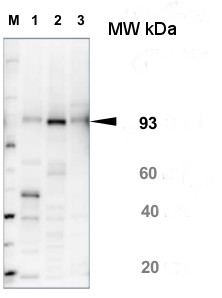
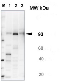
10 µg of total protein from Arabidopsis thaliana leaf (1) , Hordeum vulgare leaf (2), Zea mays leaf (3) were extracted with Protein Extraction Buffer PEB (AS08 300). Samples were diluted with 1X sample buffer (NuPAGE LDS sample buffer (Invitrogen) supplemented with 50 mM DTT and heat at 70°C for 5 min and keept on ice before loading. Protein samples were separated on 4-12% Bolt Plus gels, LDS-PAGE and blotted for 70 minutes to PVDF using tank transfer. Blots were blocked immediately following transfer in 2% blocking reagent or 5% non-fat milk dissolved in 20 mM Tris, 137 mM sodium chloride pH 7.6 with 0.1% (v/v) Tween-20 (TBS-T) for 1h at room temperature with agitation. Blots were incubated in the primary antibody at a dilution of 1: 10 000 (in blocking reagent) for 1h at room temperature with agitation. The antibody solution was decanted and the blot was rinsed briefly twice, and then washed 1x15 min and 3x5 min with TBS-T at room temperature with agitation. Blots were incubated in secondary antibody (anti-rabbit IgG horse radish peroxidase conjugated, recommended secondary antibody AS09 602, Agrisera) diluted to 1:25 000 in blocking reagent for 1h at room temperature with agitation. The blots were washed as above. The blot was developed for 5 min with detection reagent according the manufacturers instructions. Images of the blots were obtained using a CCD imager (VersaDoc MP 4000) and Quantity One software (Bio-Rad). Exposure time was 1 minute.Application examples: 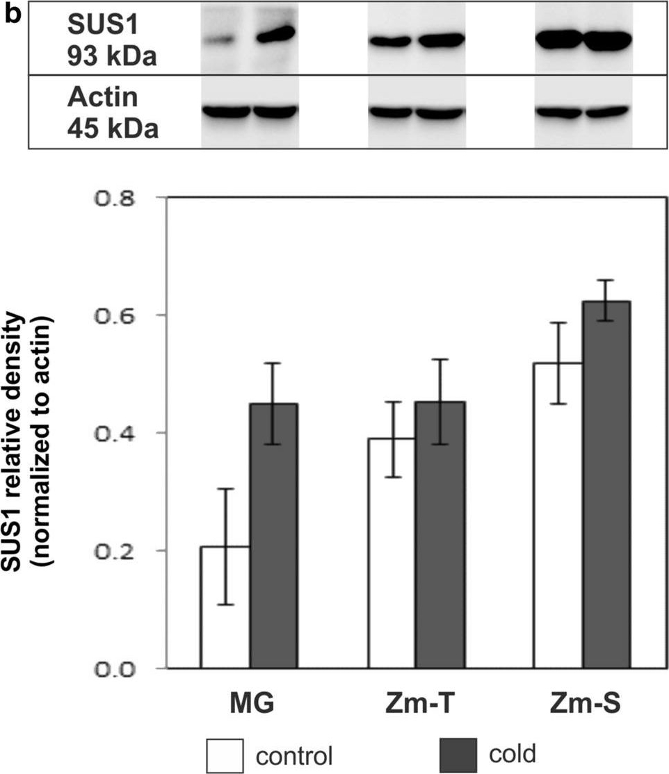
Reactant: Zea mays (Maize/Corn)
Application: Western Blotting
Pudmed ID: 32676847
Journal: Planta
Figure Number: 1B
Published Date: 2020-07-16
First Author: Bilska-Kos, A., Mytych, J., et al.
Impact Factor: 3.95
Open PublicationThe relative expression of a sucrose phosphate synthase (SPS) and b sucrose synthase 1 (SUS1) in leaves of the control (white bars) and chilled (grey bars) plants of Miscanthus?×?giganteus (MG), chilling-tolerant (Zm-T) and chilling-sensitive maize line (Zm-S). Representative immunoblots are reported. The normalization was performed relative to actin. a For SPS, there is a significant effect of genotype [ANOVA, F (2;12)?=?68.73; P?<?0.0001], treatment [ANOVA, F (1;12)?=?90.83; P?<?0.0001] and of the interaction of genotype: treatment [ANOVA, F (2;12)?=?5.14; P?=?0.024]. b For SUS1, there is a significant effect of genotype [ANOVA, F (2;12)?=?18.39; P?=?0.0002], treatment [ANOVA, F (1;12)?=?17.10; P?=?0.0014], yet there is no significant effect of the interaction of genotype: treatment [ANOVA, F (2;12)?=?2.73; P?=?0.11]. Protein lysates separated during SDS-PAGE electrophoresis were obtained by pooling the leaf material from at least 6 plants for each treatment from three independent experiments (n?=?3). Bars represent the means?±?SD, asterisks indicating a significant effect of chilling (Tukey’s HSD test): ***P???0.001
- Additional Information
-
Additional information: Antibody is recognizing recombinant SUS1 protein - Background
-
Background: SUS1 (Sucrose synthase 1) is a sucrose-cleaving enzyme that provides UDP-glucose and fructose for various metabolic pathways. Alternative names: ASUS1, ATSUS1. SUS1. - Product Citations
-
Selected references: Çetinbaş Genç et al. (2026). Tolerance mechanisms to UV-B in olive pollen demonstrate cultivar-dependent biochemical responses. Plant Physiol Biochem. 2026 Apr 2:233:111264. doi: 10.1016/j.plaphy.2026.111264.
Bilska-Kos et al. (2020). Sucrose phosphate synthase (SPS), sucrose synthase (SUS) and their products in the leaves of Miscanthus× giganteus and Zea mays at low temperature. Planta . 2020 Jul 16;252(2):23. doi: 10.1007/s00425-020-03421-2.
Kleczkowski LA & Decker DD (2015) Sugar activation for production of nucleotide sugars as substrates for glycosyltransferases in plants. J. Appl. Glycosci. (in press). - Protocols
- Agrisera Western Blot protocol and video tutorials
- Reviews:
-
This product doesn't have any reviews.
Related products

AS09 602 | Clonality: Polyclonal | Host: Goat | Reactivity: Rabbit IgG (H&L)