Visar bild 1 av
1

Anti-5-mC | 5-methylcystosine (monclonal)
Product no: AS16 3161
AS16 3161 | Clonality: monoclonal | Host: Mouse | Reactivity:Human, Mouse, other species
427
€
Customer reviews
Delivery:
3-6 business days
- Product Info
-
Immunogen: BSA-conjugated molecule: 5-methylcytosine (5-mC) Sub class: IgG1 Host: Mouse Clonality: Monoclonal Purity: IgG Protein A purified. Format: Liquid Quantity: 50 µg Storage: Store lyophilized/reconstituted at -20°C; once reconstituted make aliquots to avoid repeated freeze-thaw cycles. Please remember to spin the tubes briefly prior to opening them to avoid any losses that might occur from material adhering to the cap or sides of the tube. Tested applications: Dot blot (Dot), Immunofluorescence (IF), immunoprecipitation (IP), FISH (FISH), MeDIP (methylated DNA Immunoprecipitation (IP) Recommended dilution: 1 : 600 (Dot), 1-5 µg (IP), 1 : 500 (IF), 1 : 200-1 : 1000 (FISH) - Reactivity
-
Confirmed reactivity: Human Predicted reactivity: Mouse, broad species range Not reactive in: No confirmed exceptions from predicted reactivity are currently known - Application Examples
-
application example 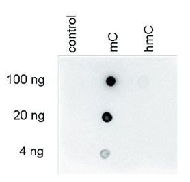
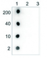
Dot blot: to demonstrate the specificity of the monoclonal antibody against 5-mC, a Dot blot analysis was performed using the hmC, mC and C controls: “5-hmC, 5-mC & cytosine DNA. One hundred to 4 ng (equivalent of 5 to 0.2 pmol of C-bases) of the controls were spotted on a membrane (Amersham Hybond-N+). The antibody was used at a dilution of 1:600.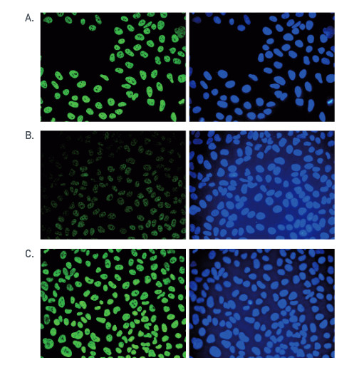
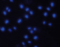
Immunofluorescence: human osteosarcoma (U2OS) cells were stained with the monoclonal antibody against 5-mC. Cells were fixed with 2.5% PFA in PBS for 30’, permeabilised with 0.5% Triton X-100 for 1 hour and treated with 2N HCl for 1 hour followed by 2 x 5 minutes with 0.1 M borate buffer to depurinate the DNA. After blocking with PBS containing 0.1% Triton X-100 and 1% BSA, the cells were immunofluorescently labelled with the 5-mC antibody diluted 1:500 in blocking solution, followed by a goat anti-mouse antibody conjugated to Alexa488. Panel (A): cells were immunofluorescently labelled with the 5-mC antibody (left) or with DAPI (right). Panel (B) and (C): staining of the cells with the 5-mC antibody after incubation of the antibody with 50 μM mCTP or hmCTP, respectively.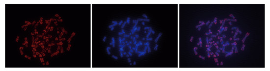
FISH: to detect methylated chromosomal regions, FISH was performed on metaphase chromosomes from HeLa cells using the monoclonal antibody against 5-mC. The cells were blocked in metaphase by treatment with colcemid (0.1 μg/ml) for 1 - 2 hours, fixed overnight at -20°C with ethanol/glacial acetic acid and treated with 2N HCl for 30’ at room temperature. Subsequently, the cells were blocked with PBS containing 1% BSA and 0.1% Triton X-100 and stained with the 5-mC antibody (left) diluted 1:1,000 in blocking solution, followed by an anti-mouse antibody conjugated to Alexa594. The middle panel shows staining of the chromosomes with DAPI. A merge of the two staining is shown to the right. - Additional Information
-
Additional information: This antibody has been purified using Protein A and is present in PBS - Background
-
Background: 5-mC (5-methylcystosine) is a methylated form of the DNA base cytosine. Methylation may be involved in the regulation of gene transcription.
Alternative names: 5 Me citidine, 5 Methycytosine, 5 Me Cytidine, methyl CpG. - Reviews:
-
This product doesn't have any reviews.
Related products
AS09 627 | Clonality: Polyclonal | Host: Rabbit | Reactivity: Mouse IgG (H&L)
225 €
AS16 3162 | Clonality: monoclonal | Host: Mouse | Reactivity:Human, Mouse, other species
427 €
AS16 3164 | Clonality: monoclonal | Host: Mouse | Reactivity:Human, Mouse, other species
544 €