1
Anti-DPE2 | 4-alpha-glucanotransferase
AS23 4952 | Clonality: Polyclonal | Host: Rabbit | Reactivity: Arabidopsis thaliana
- Product Info
-
Immunogen: KLH-conjugated peptide derived from Arabidopsis thaliana DPE2 protein sequence, UniProt: Q8RXD9 TAIR: AT2G40840 Host: Rabbit Clonality: Polyclonal Purity: Antigen affinity purified serum, in PBS pH 7.4 Format: Lyophilized Quantity: 50 µg Reconstitution: For reconstitution, add 50 µl of sterile or deionized water. Storage: Store lyophilized/reconstituted at -20°C; once reconstituted, make aliquots to avoid repeated freeze-thaw cycles. Please, remember to spin tubes briefly prior to opening them to avoid any losses that might occur from lyophilized material adhering to the cap or sides of the tubes. Tested applications: Western blot (WB) Recommended dilution: 1: 500 - 1 : 1000 (WB) Expected | apparent MW: 107.7 kDa - Reactivity
-
Confirmed reactivity: Arabidopsis thaliana Predicted reactivity: Arachis hypogaea, Brachypodium distachyon, Brassica napus, Cannabis sativa, Glycine max, Gossypium sp., Hordeum vulgare, Malus domestica, Manihot esculenta, Medicago truncatula, Nicotiana tabacum, Oryza sativa, Pisum sativum, Populus sp., Ricinus communis, Solanum lycopersicum, Solanum tuberosum, Sorghum bicolor, Spinacia oleracea, Theobroma cacao, Triticum sp., Vitis vinifera, Zea mays
Species of your interest not listed? Contact usNot reactive in: No confirmed exceptions from predicted reactivity are currently known - Application Examples
-

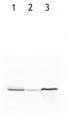
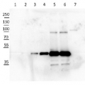
Samples:
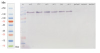
1 - Protein Ladder
2-4 - Columbia-0, Arabidopsis thaliana wild type
5-7 - fzl-3 mutant (overexpressor)
8-10 - dpe2-5/phs1b mutant (knockout)Proteins were extracted from Arabidopsis thaliana leaves by harvesting fresh tissue into 1.5-mL microcentrifuge tubes, immediately freezing the samples in liquid nitrogen, and adding 100–200 μL of ice-cold Extraction buffer (100 mM HEPES/NaOH, pH 7.5, 5 mM DTE, 10% [m/v] glycerol, 1 mM EDTA, 200 μM PMSF). Samples were vortexed thoroughly, centrifuged at 14,000 rpm for 15 min at 4 °C, and the resulting supernatants were transferred into fresh pre-chilled tubes. All steps were carried out on ice. Protein concentrations were determined using the Bradford assay, and crude extracts were used immediately or stored at –80 °C.
For SDS–PAGE analysis, 40 μg of total protein was mixed with 3× SDS sample buffer (final concentration 62,5 mM Tris/HCl pH 6.8, 10% [w/v] glycerol, 2% [w/v] SDS, 20 mM DTE, 0.007% [w/v] Bromophenol blue), heated at 95 °C for 5 min at 1200 rpm, and centrifuged at room temperature for 15 min at 14,000 × g. The components of the SDS–PAGE gels were as follows a 9% separating gel was prepared using 6 mL 0.75 M Tris–HCl (pH 8.8), 120 μL 10% SDS, 3.6 mL Rotiphorese® GEL 30, 40 μL 10% ammonium persulfate, 6 μL TEMED, and 3 mL distilled water. The stacking gel was prepared using 2 mL 0.5 M Tris–HCl (pH 6.8), 80 μL 10% SDS, 0.8 mL Rotiphorese® GEL 30, 80 μL 10% ammonium persulfate, 6 μL TEMED, and 5 mL distilled water. The running buffer consisted of 30.3 g Tris, 144.12 g glycine, and 10 g SDS made up to 1 L with distilled water.
For western blotting, proteins were transferred using cold transfer buffer (50 mM Tris, 150 mM glycine, 20% [v/v] methanol; and 0.02% [w/v] SDS) at a constant voltage of 40 V for 50 min. with current set to unlimited (maximum 250 mA). Transfers were performed on ice using a wet-transfer system with nitrocellulose membranes (0.2 μm pore size). Membranes were blocked with 5% (w/v) milk in TBS-T for 1 h at room temperature with gentle agitation. The blot was incubated with primary antibody diluted 1:500 in 5% milk-blotting buffer or 1 h at room temperature with gentle agitation. After incubation, the antibody solution was discarded, and the membrane was washed six times for 5 min each in TBS-T. The blot was then incubated with secondary antibody (anti-rabbit IgG, horse alkaline-phosphatase conjugated; Agrisera AS09 607) diluted 1:1000 for 30 min to 1 h at room temperature with gentle agitation. Following again six washes in TBS-T (5 min each).
For signal development, the membrane was incubated in alkaline phosphatase (AP) buffer supplemented with NBT and BCIP until an appropriate signal intensity was obtained. For each membrane, 15 mL of AP buffer (100 mM Tris-HCl, pH 9.5, 100 mM NaCl, 5 mM MgCl₂) was mixed with 99 μL NBT solution (50 mg NBT dissolved in 1 mL 70% [v/v] DMF) and 49.5 μL BCIP solution (50 mg BCIP dissolved in 1 mL DMF). The membrane was incubated in this developing solution at room temperature until the desired signal appeared.
Courtesy of Biopolymer Analytics, University of Potsdam, Germany - Background
-
Background: DPE2 (4-alpha-glucanotransferase) is a cytosolic alpha-glucanotransferase essential for the cytosolic metabolism of maltose, an intermediate on the pathway by which starch is converted to sucrose in leaves at night. This enzyme is metabolizing maltose exported from the chloroplast and is specific for beta-maltose. May play a role in freezing tolerance. - Protocols
-
Agrisera Western Blot protocol and video tutorials
Protocols to work with plant and algal protein extracts
Agrisera Educational Poster Collection - Reviews:
-
This product doesn't have any reviews.
Related products

AS09 602 | Clonality: Polyclonal | Host: Goat | Reactivity: Rabbit IgG (H&L)

AS09 607 | Clonality: Polyclonal Host: Goat Reactivity: Rabbit IgG (H&L)
AS11 1739 | Clonality: Polyclonal | Host: Rabbit | Reactivity: A. thaliana, C. reinhardtii, H. vulgare, N. tabacum, Polytomella sp., T. aestivum, Z. mays
AS09 462 | Clonality: Polyclonal | Host: Rabbit | Reactivity: Arabidopsis thaliana
AS04 043 | Clonality: Polyclonal | Host: Rabbit | Reactivity: Arabidopsis thaliana, Brassica napus, Macroptilium atropurpureum, Nicotiana benthamiana, Pinus silvestris, Pinus yunanniensis, Oryza sativa, Petunia hybrida cv. Mitchell, Solanum tuberosum, Zea mays | cellular [compartment marker] of cytoplasm