1
Anti-RFP | Red flourescent protein (mRFP, mCherry, tdTomato, mScarlet)
AS24 5006 | Clonality: Polyclonal | Host: Rabbit | Reactivity: mRFP/mCherry/tdTomato/mScarlet tagged proteins
- Product Info
-
Immunogen: Recombinant protein produced in E.coli corresponding to full lenght mRFP. The sequence used for immunization is also found in other red flourescent proteins like: mCherry/tdTomato/mScarlet and others. Host: Rabbit Clonality: Polyclonal Purity: Antigen affinity purified serum, in PBS pH 7.4 Format: Lyophilized Quantity: 50 µg Reconstitution: For reconstitution, add 50 µl of sterile or deionized water. Storage: Store lyophilized/reconstituted at -20°C; once reconstituted make aliquots to avoid repeated freeze-thaw cycles. Please, remember to spin tubes briefly prior to opening them to avoid any losses that might occur from lyophilized material adhering to the cap or sides of the tubes. Tested applications: Immunohistochemistry (IHC), Western blot (WB) Recommended dilution: 1: 200 (IHC), 1: 2500 (WB) Expected | apparent MW: Depends upon fusion partner - Reactivity
-
Confirmed reactivity: RFP, mCherry-YFP, mScarlet Predicted reactivity: mRFP, mCherry, tdTomato Not reactive in: Sequence identity with GFP is very low and it is therefore expected that this antibody does not cross-reacti with GFP protein. - Application Examples
-
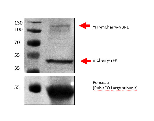
Samples:
1 – 1 ul PageRuler™ Plus Prestained Protein Ladder, 10 to 250 kDa (ThermoFischer Scientific #26619)
2 – 20 ul of Arabidopsis thaliana YFP-mCherry-NBR1 5 week old (6 leaf discs (12,56 mm2) pulverized in 100ul 1X SDS-PAGE gel loading buffer).
6 leaf discs (12,56 mm2) were pulverized and denatured in 100 ul 1X SDS-PAGE gel loading buffer at 95 °C 5 min following Svenning et al. 2011 protocol (doi: 10.4161/auto.7.9.16389). 20 µl were loaded and separated on 15% SDS-PAGE and blotted for 1 h to PVDF using wet transfer in the cold. Blot was blocked with 5% milk for: 1h/RT with agitation. Blot was incubated in the primary antibody at a dilution of 1:1000 in TBS-T ON/4°C with agitation. The antibody solution was decanted, and the blot was washed 3 times for 5 min in TBS-T at RT with agitation. Blot was incubated in matching secondary antibody (anti-rabbit IgG horse radish peroxidase conjugated) diluted to 10 000 in for 2h/RT with agitation. The blot was washed 2 times for 5 min in TBS-T, 2 times for 5 min in TBS and incubated for 5 min in HRP buffer (100 mM Tris-HCl pH9.5, 5mM MgCl2, 100 mM NaCl) prior incubation with HRP buffer supplied with 0.15 mg/ml HRP and 0.33 mg/ml NBT for 20 min at RT with agitation. Membrane picture was taken with a GelDoc Go Imaging System (Bio-rad).
Courtesy of Dr. Ignacio Lescano López, Centro de Investigaciones Agropecuarias (INTA-CIAP), Argentina

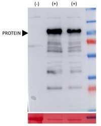
Samples, from left to right:
1 - 50 ug of 7 day-old Arabidopsis thaliana seedlings (cotyledons, leaves, hypocotyl and root)
2 - 5 ug of Arabidopsis thaliana expressing the protein of interest fused to RFP
3 - 5 µg of Arabidopsis thaliana expressing the protein of interest fused to red fluorescent protein (RFP) and another protein fused to green fluorescent protein (GFP).
1 - 50
5 μg/well of total protein extracted freshly from 7-day-old Arabidopsis thaliana seedlings. Exact buffer components were: 25 mM Tris-HCl pH 7.5, 10% glycerol, 1 mM EDTA pH 8.0, 150 mM NaCl, 10 mM DTT, and 1x protease inhibitor cocktail [cOmplete, EDTA-free; Roche] and denatured with Laemmli buffer 100°C during 5 min. Samples were separated in the cold on 10 % SDS-PAGE and blotted for 1h to PVDF, using wet transfer in the cold. Blot was blocked with 5% milk for: 1h/RT with agitation. Blot was incubated in the primary antibody at a dilution of 1: 2500 for 1hr at room temperature with agitation. The antibody solution was decanted and the blot was rinsed briefly twice, then washed once for 15 min and 3 times for 5 min in TBS-T at RT with agitation. Blot was incubated in matching secondary antibody anti-rabbit IgG horse radish peroxidase conjugated, AS09 602, Agrisera) diluted to 1:25 000 for h/RT with agitation. The blot was washed as above and developed with a low femtogram chemiluminescent detection reagent. To decrease the background signal further, less sensitive detection reagent can be applied.Courtesy of Dr. Victoria Gastaldi, Institute of Molecular and Cellular Biology of Plants (IBMCP, UPV-CSIC), Valencia, Spain
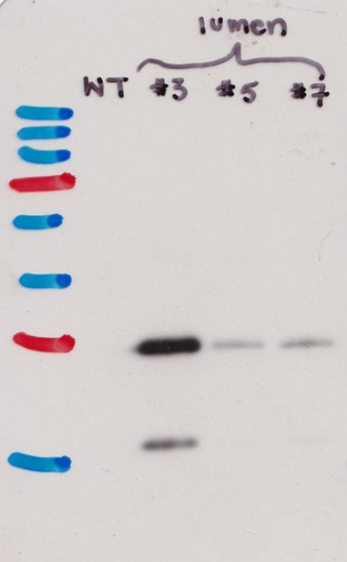
Samples:
The first lane from the left: Physcomitrium patens WT
# 3, 5, 7 Physcomitrium patens lines overexpressing mScarlet3 tag.
Protein extraction: moss (Physcomitrium patens) tissue was ground into powder with liquid nitrogen in a mortar and pestle. The frozen tissue powder was directly mixed with 2x Laemmli sample buffer with freshly added β-mercaptoethanol). The samples were denatured at 95°C for 5 min, cooled on ice, and spun down. The supernatant was loaded on the gel. Samples were separated in 10% SDS-PAGE (with 4% stacking) and blotted for 1h to PVDF membrane, using wet transfer in the cold. Blot was blocked with 5% Milk at 4°C/ON with agitation. Blot was incubated in the primary antibody at a dilution of 1:2 500 for 1h/RT with agitation in TBS-T with 5% milk. The antibody solution was decanted, and the blot was rinsed briefly, then washed three times for 5 min in TBS-T at RT with agitation. Blot was incubated in matching secondary antibody (anti-rabbit IgG horse radish peroxidase conjugated) diluted to 1:25 000 in TBS-T with 5% milk and incubated for 1h/RT with agitation. The blot was washed as above, but with 10 min wash steps, and developed with the following chemiluminescent detection reagent.
Courtesy of Dr. Ivan Radin, University of Minnesota, USA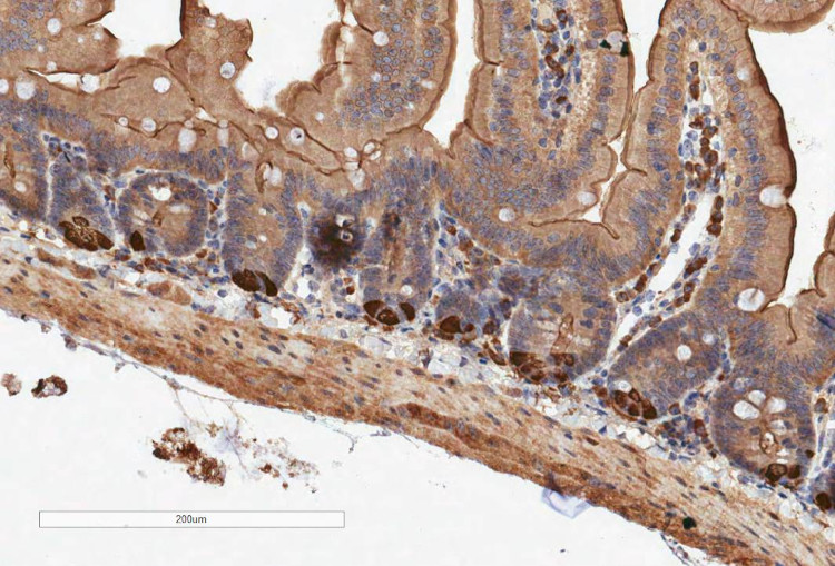
Type of material: Mouse intestinal tissue (FFPE tissue), overexpressing mScarlet tagged protein
Fixation: formaldehyde
Hydrophilization: n/a
Cell wall digestion: n/a
Membrane permeabilization: n/a
Antigen retrieval: in citrate buffer
Blocking buffer: Superblock plus (Thermoscientific 37580) Washing buffer: PBS
Primary antibody dilution and incubation time: 1:200/ 1 h incubation
Secondary antibody: Vector labs, BA-1000-Goat Anti-Rabbit IgG Antibody (H+L), Biotinylated 1:200
Co-staining of the nucleus: HaematoxylinCourtesy of Dr. Hayley Belnoue-Davis, Centre for Human Genetics, University of Oxford, United Kingdom
- Background
-
Background: Red fluorescent protein (RFP) once excited is fluorescing red-orange light. The mass of RFP is approximately 25.9 kDa and its excitation maximum is 558 nm and emission maximum is 583 nm. Improved variants of RFP include so called mFruits variants: mCherry, mOrange, mRaspberry. - Product Citations
-
Selected references: 38221900 - Protocols
-
Agrisera Western Blot protocol and video tutorials
Protocols to work with plant and algal protein extracts
Agrisera Educational Poster Collection - Reviews:
-
This product doesn't have any reviews.
Related products

AS09 602 | Clonality: Polyclonal | Host: Goat | Reactivity: Rabbit IgG (H&L)

AS20 4485 | Clonality: Monoclonal | Host: Mouse | Reactivity: BFP, GFP, YFP
AS11 1775 | Clonality: Polyclonal | Host: Rabbit | Reactivity:C-terminal of YFP
AS20 4512 | Clonality: Polyclonal | Host: Rabbit | Reactivity: FP, YFP, EGFP | Green Fluorescence Protein and its variants