1
Anti-DnaK | chloroplast stromal chaperone
AS07 270 | Clonality: Polyclonal | Host: Rabbit | Reactivity: Chlamydomonas reinhardtii, Synechocystis 6803 motile, Synechocystis 6803 GT (glucose tolerant strain), Synechococcus elongates 7942
- Product Info
-
Immunogen: recombiant HSP70B of Chlamydomonas reinhardtii (XP_001696432), UniProt: A8HYV3
Host: Rabbit Clonality: Polyclonal Purity: Serum Format: Lyophilized Quantity: 50 µl Reconstitution: For reconstitution add 50 µl of sterile water Storage: Store lyophilized/reconstituted at -20°C; once reconstituted make aliquots to avoid repeated freeze-thaw cycles. Please remember to spin the tubes briefly prior to opening them to avoid any losses that might occur from material adhering to the cap or sides of the tube. Tested applications: Western blot (WB) Recommended dilution: 1 : 5000 (WB) Expected | apparent MW: 70 kDa
- Reactivity
-
Confirmed reactivity: Chlamydomonas reinhardtii, Synechocystis 6803 motile, Synechocystis 6803 GT (glucose tolerant strain), Synechococcus elongates sp. PCC7942 Predicted reactivity: Species of your interest not listed? Contact us Not reactive in: No confirmed exceptions from predicted reactivity are currently known - Application Examples
-
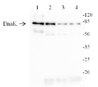
Application example 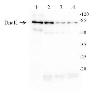
15 µg of Arabidopsis thaliana leaf extract (1), 10 µg of total protein from: Synechocystis 6803 motile (2), Synechocystis 6803 GT (glucose tolerant strain) (3), Synechococcus elongates 7942 (4), Marker - P ierce™ Prestained Protein MW Marker (kat #26612): Total protein was extracted with following buffer: 10 mM Tris HC l, pH 8.0, 0.5% LDS, 4% glycerol, 0.1 mM EDTA were mixed with sample buffer and denatured for 5 min at 95°C. Samples were separated on 10% S DS -PAGE a nd b lo tted 1 h to nitrocellulose membrane (Amersha m Protran) using tank wet transfer (Bio -Rad) in standard transfer buffer in presence of 20% methanol. Transfer of proteins to the membrane was checked using 0,5% Ponceau S staining before the blocking step. Blots were blocked in buffer (2 % lo w -fat milk in 1xPBS, 0,1% Tween) for 1 h at room temperature (RT) with agitation. Blots were incubated in the primary antibody at a dilution of 1 : 5000 for 1 h at RT with agitation. The antibody solutionwas decanted and the blot was rinsed briefly twice, then washed once f or 15 min and 3 times for 5 min in PBS -T at RT with agitation. Blot was incubated in secondary antibody (goat anti-rabbit IgG, AS09 602, Agrisera ) dilut ed to 1 :30 000 in for 1 h at RT with agitation. The blot was was washed as above and developed for 5 min with Clarity Western ECL Substrate and ChemiDoc detection system (Bio-Rad).
Courtesy Dr. Elena Pojidaeva, Laboratory of Plant Gene Expression, Timiryazev Institute of Plant Physiology RAS, 127276 Moscow Russia - Additional Information
-
Additional information (application): It is not determined which isoform of DnaK is recognized by this antibody in Arabidopsis thaliana. - Background
-
Background: Procaryotic Hsp70 protein family includes DnaK, HscA (Hsc66), HscC (Hsc62). Those proteins are involved in protein folding, cell protection from environmental stress and other functions which are vital for a living cell.
- Product Citations
-
Selected references: Göhre et al. (2006). One of Two Alb3 Proteins Is Essential for the Assembly of the Photosystems and for Cell Survival in Chlamydomonas The Plant Cell 18:1454–1466. - Protocols
-
Agrisera Western Blot protocol and video tutorials
Protocols to work with plant and algal protein extracts
Oxygenic photosynthesis poster by prof. Govindjee and Dr. Shevela
Z-scheme of photosynthetic electron transport by prof. Govindjee and Dr. Björn and Dr. Shevela - Reviews:
-
This product doesn't have any reviews.
Related products
AS07 271 | Clonality: Polyclonal | Host: Rabbit | Reactivity: Chlamydomonas reinhardtii

AS09 607 | Clonality: Polyclonal Host: Goat Reactivity: Rabbit IgG (H&L)

AS09 602 | Clonality: Polyclonal | Host: Goat | Reactivity: Rabbit IgG (H&L)