1
Anti-Gamma CA | Gamma Carbonic anhydrases
AS17 4114 | Clonality: Polyclonal | Host: Rabbit | Reactivity: Arabidopsis thaliana, Oryza sativa
- Product Info
-
Immunogen: KLH-conjugated peptide derived from Arabidopsis thaliana Gamma-Carbonic anhydrase, UniProt: Q9C6B3, TAIR: AT1G47260
Host: Rabbit Clonality: Polyclonal Purity: Serum Format: Lyophilized Quantity: 50 µl Reconstitution: For reconstitution add 50 µl of sterile water, Serum contains 0,1 % sodium azide as preservative Storage: Store lyophilized/reconstituted at -20°C; once reconstituted make aliquots to avoid repeated freeze-thaw cycles. Please remember to spin the tubes briefly prior to opening them to avoid any losses that might occur from material adhering to the cap or sides of the tube. Tested applications: Western blot (WB) Recommended dilution: 1 : 1000 (WB) Expected | apparent MW: 9.97 KDa (gamma-CA1), 30KDa (gamma-CA2) strongest band, 27.83 (gamma-CA3), 25.08 KDa Arabidopsis thaliana proteins - Reactivity
-
Confirmed reactivity: Arabidopsis thaliana, Oryza sativa Predicted reactivity: Arachis duranensis, Brachypodium distachyon, Brassica oleracea, Brassica rapa, Camelina sativa, Capsella rubella, Citrus sinensis, Daucus carota, Erythranthe guttatus, Fragaria vesca subsp. vesca, Gossypium hirsutum, Jatropha curcas, Juglans regia, Lupinus angustifolius, Malus x domestica, Medicago truncatula, Nelumbo nucifera, Oryza brachyantha, Populus euphratica, Prunus mume, Pyrus x bretschneideri, Raphanus sativus, Sesamum indicum, Setaria italica, Solanum tuberosum, Tarenaya hassleriana, Theobroma cacao, Triticum aestivum, Zea mays, Zostera muelleri
Species of your interest not listed? Contact us - Application Examples
-
application example 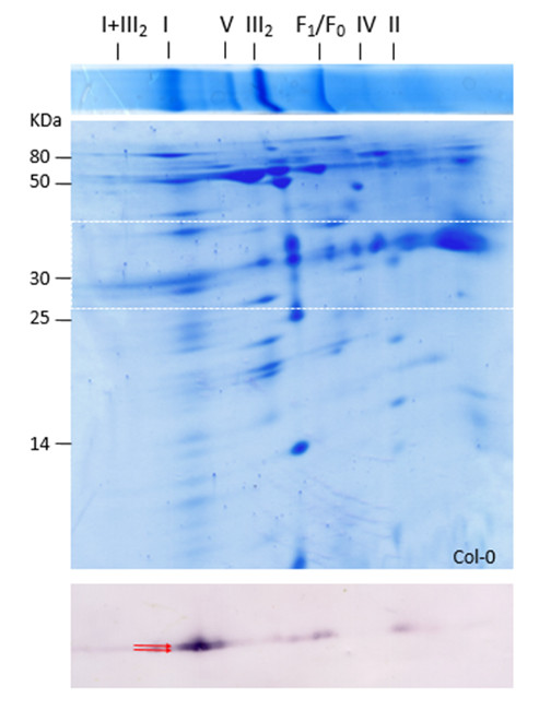
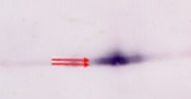
Mitochondrial proteins (1mg) from Arabidopsis thaliana ecotype Col-0 cell suspensions were separated by 2D BN/SDS-PAGE (Klodmann et al., 2011; Plant Physiology 157: 587-598). After gel electrophoresis, proteins were transferred to nitrocellulose membrane using semi-dry conditions (90 min; 0.8mA per cm2 of gel). After washing once in TBS-T buffer (10 min), blot was blocked overnight at 4ºC with 10% p/v milk in TBS-T. Blot was washed three times with 0.5% p/v milk in TBS-T (10 min each) and incubated 1 hour with primary antibody (1:1000 in TBS-T, 3% BSA and 0.02% NaN3). Washing was carried out three times with TBS-T, 0.5% BSA, 10 min each. Then blot was incubated 1 hour with secondary antibody (Anti-rabbit IgG conjugated to alkaline phosphatase at a dilution of 1:10 000) in TBS-T, 3% BSA, 0.02% NaN3). After washing (as above), blot was equilibrated 5 minutes in AP buffer (Tris-HCl 1M pH 9.5; 5M NaCl; 4M MgCl2). Finally, revealing was developed by incubation in AP buffer; 110 µg/ml NBT; 75 µg/ml BCiP. Reaction was stopped by discarding revealing solution and adding distilled water. Upper lane: 1D BN gel electrophoresis of mitochondrial proteins, with respective complexes.
Lower gel: second dimension, where mitochondrial proteins of complexes were separated. Blot section (indicated as dashed square in gel) reveals mainly two bands (around 30 kDa- arrows), corresponding to both gamma-CA2 forms. The smear along the 30 kDa line is interpreted as fragments of complex I containing CA2 protein (Perales et al., 2005; Journal of Molecular Biology, 350: 263-277).Application examples: 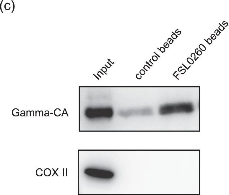
Reactant: Arabidopsis thaliana (Thale cress)
Application: Western Blotting
Pudmed ID: 32457324
Journal: Sci Rep
Figure Number: 4C
Published Date: 2020-05-26
First Author: Sako, K., Futamura, Y., et al.
Impact Factor: 4.13
Open PublicationFSL0260 interacts with mitochondrial complex I and inhibits its activity. (a) Inhibition of complex I by FSL0260. Oxygen consumption rate (OCR) of isolated mitochondria from potato tuber was monitored with deamino-NADH in the absence or presence of FSL0260. (b) Inhibition of deamino-NADH oxidation by FSL0260. Deamino-NADH oxidation was measured by spectrometry using sonicated mitochondria isolated from the potato tuber. The experiment was conducted using three biological replicates. Error bars represent the mean ± SE (n?=?3). (c) Pull-down assay of FLS0260. Sonicated potato mitochondria were incubated with control or FSL0260 beads. Immunoblot assay was performed using an anti-gamma CA antibody and an anti-COX II antibody.
- Additional Information
-
Additional information: Contains 0,1% ProClin - Background
-
Background: Gamma CAH (Gamma Carbonic anhydrase) proteins are localized in the inner mitochondrial membrane. They are in a range of 25-30 kDa.
- Product Citations
-
Selected references: Chen et al. (2019). Composition of Mitochondrial Complex I during the Critical Node of Seed Aging in Oryza sativa. Journal of Plant Physiology Volume 236, May 2019, Pages 7-14.
Kühn et al. (2015). Complete Mitochondrial Complex I Deficiency Induces an Up-Regulation of Respiratory Fluxes That Is Abolished by Traces of Functional Complex I. Plant Physiol. 2015 Aug;168(4):1537-49. doi: 10.1104/pp.15.00589. Epub 2015 Jul 1. - Protocols
-
Agrisera Western Blot protocol and video tutorials
Protocols to work with plant and algal protein extracts
Oxygenic photosynthesis poster by prof. Govindjee and Dr. Shevela
Z-scheme of photosynthetic electron transport by prof. Govindjee and Dr. Björn and Dr. Shevela - Reviews:
-
This product doesn't have any reviews.
Related products

AS09 607 | Clonality: Polyclonal Host: Goat Reactivity: Rabbit IgG (H&L)

AS09 602 | Clonality: Polyclonal | Host: Goat | Reactivity: Rabbit IgG (H&L)
AS05 073 | Clonality: Polyclonal | Host: Rabbit | Reactivity: Chlamydomonas reinhardtii