1
Anti-GFP | Green Fluorescence Protein
AS20 4443 | Clonality: Polyclonal | Host: Rabbit | Reactivity: Green Fluorescence Protein (GFP)
- Product Info
-
Immunogen: Recombinant GFP protein derived from Aequorea victoria, UniProt: P42212
Host: Rabbit
Clonality: Polyclonal Purity: Immunogen affinity purified serum in PBS pH 7.4. Format: Lyophilized Quantity: 50 µg Reconstitution: For reconstitution add 50 µl of sterile water Storage: Store lyophilized/reconstituted at -20°C; once reconstituted make aliquots to avoid repeated freeze-thaw cycles. Please remember to spin the tubes briefly prior to opening them to avoid any losses that might occur from material adhering to the cap or sides of the tube. Tested applications: Chromatin Immunoprecipitation (ChIP), Immunoprecipitation (IP), Western blot (WB) Recommended dilution: 1: 5000 - 1 : 10 000 (WB) - Reactivity
-
Confirmed reactivity: Recombinant GFP overexpressed in E.coli from Arabidopsis thaliana, Chlamydomonas reinhardii, Nicotiana tabacum Predicted reactivity: mClover3 - Application Examples
-
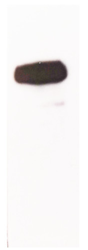
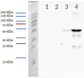
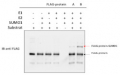
19.4 µg/well of total protein extracted freshly from tobacco with buffer (50mM Tris pH 7.5, 150 mM NaCl, 1 mM EDTA, 10% Glicerol, 1 mM DTT, 1X Pefablock) and denatured with SDS 2% and 0.1% Bromophenol blue (Laemmli buffer) at 95°C for 5 min. Separated on 12 % SDS-PAGE and blotted 2h to PVDF/nitrocellulose, using wet transfer. Blot was blocked with 5% milk for 1h/RT with agitation. Blot was incubated in the primary antibody at a dilution of 1: 10 000 in TBS-T ON/4°C with agitation. The antibody solution was decanted and the blot was rinsed briefly twice, then washed 3 times for 10 min in TBS-T at RT with agitation. Blot was incubated in Agrisera matching secondary antibody (anti-rabbit IgG horse radish peroxidase conjugated, AS09 602) diluted to 1:20 000 in for 1h/RT with agitation. The blot was washed as above and developed for 2 min with Agrisera ECLSuperBright. Exposure time was 60 seconds.Courtesy of Delfina Gagliardi Instituto de Agrobiotecnología del Litoral (IAL), Argentina
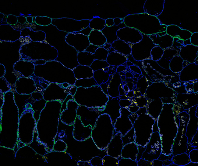
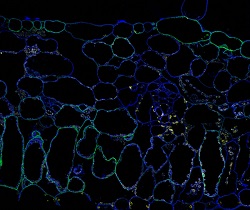
Super-resolution image of immunofluorescent localization of GFP-fused protein in Nicotiana tabacum (tobacco) leaf tissue using Agrisera anti-GFP antibodies (AS20 4443) and donkey anti-rabbit IgG DyLight 650 conjugated secondary antibodies (Green). Calcofluor White (Blue) and Tissue Autofluorescence (Yellow),Method:
Fixation: 2% paraformaldehyde + 0.5% glutaraldehyde, dehydration in ethanol, embedment and UV polymerization in HM20 resin
Cell wall digestion: no
Membrane permeabilization: DMSO-IGEPA
Air drying; No
Antigen retrieval: No
Blocking buffer: 4% w/v non-fat milk powder in 1X Phosphate Buffered Saline buffer, prepared freshly, filtered with 0.2 µm filer
Washing buffer: 1X Phosphate Buffered Saline buffer Primary antibody dilution and incubation time: 1:100; 1 hour at room temperature
Secondary antibody dilution and incubation time and supplier: 1:200 Donkey anti-rabbit IgG (H&L) DyLight®650 (Agrisera, AS12 2254); 1 hour at room temperature
Co-staining of the nucleus (DAPI): No
Cell wall and nucleus staining: Calcofluor White
All material was freshly prepared. All incubation volumes were kept the same: 200 μL per 22x22 mm cover glass/sample
Courtesy of Dr. Kirk Czymmek, Donald Danforth Plant Science Centre, USA. - Background
-
Background: GFP (Green fluorescent protein) was originally identified in photo organs on jellyfish Aequorea victoria. It is a naturally fluorescent protein which emits green light at a maximum wavelength of 509 nm when excited by blue or UV light. It is extensively used in laboratory as a reporter molecule to label and study cellular and subcellular proteins in living cells using a wide range of applications. Antibodies to GFP protein are used in immunoblotting and ELISA. GFP protein has molecular weight of 27 kDa. - Protocols
-
Agrisera Western Blot protocol and video tutorials
Protocols to work with plant and algal protein extracts
Agrisera Educational Poster Collection - Reviews:
-
This product doesn't have any reviews.
Related products

AS09 602 | Clonality: Polyclonal | Host: Goat | Reactivity: Rabbit IgG (H&L)
AS20 4441 | Clonality: Polyclonal | Host: Rabbit | Reactivity: 3xHis-tag
AS20 4442 | Clonality: Polyclonal | Host: Rabbit | Reactivity: DYKDDDDK-Tag