1
Anti-His tag | 3xHis (polyclonal)
AS20 4441 | Clonality: Polyclonal | Host: Rabbit | Reactivity: 3xHis-tag
- Product Info
-
Immunogen: KLH-conjugated synthetic peptide 3xHis
Host: Rabbit Clonality: Polyclonal Purity: Antigen affinity purified serum in PBS pH 7.4. Format: Lyophilized Quantity: 50 µg Reconstitution: For reconstitution add 50 µl, of sterile water Storage: Store lyophilized/reconstituted at -20°C; once reconstituted make aliquots to avoid repeated freeze-thaw cycles. Please remember to spin the tubes briefly prior to opening them to avoid any losses that might occur from material adhering to the cap or sides of the tube. Tested applications: Immunofluorescence (IF), Western blot (WB) Recommended dilution: 1 µg/mL (IF), 1: 1000 - 1 : 5000 (WB) Expected | apparent MW: 3xHis tagged proteins - Reactivity
-
Confirmed reactivity: 3xHis tagged proteins - Application Examples
-
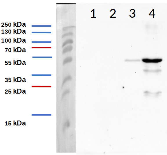
Samples:
40 µg of E. coli supernatant in control conditions at 3h (1)
40 µg of E. coli pellet in control conditions at 3h (2)
40 µg of E. coli supernatant induced at 3h (3)
40 µg of E. coli pellet induced at 3h (4)
40 µg of total protein from Escherichia coli expressing pGEX-PEX11a inducible by IPTG, extracted with HEPES 50 mM pH 7.6, 300 mM NaCl, 5% glycerol, 20 mM imidazole, 1 mM MgCl2, 1 x protease inhibitor; prepared in 0.063 M Tris-HCl buffer, pH 6.8, containing 2% sodium dodecyl sulfate (SDS; w/v), 10 % glycerol (v/v), 0.006 % bromophenol blue (w/v) and 10 mM DTT; and denaturated at 95 ºC for 5 min. Proteins were separated on 12 % SDS-PAGE and blotted 1 h to PVDF using semi-dry transfer. Blots were blocked with 3% (w/v) skimmed milk powder diluted in TBS-T O/N at 4 ºC with agitation. Blot was incubated in the primary antibody at a dilution of 1: 5.000 for 1 h at RT with agitation in TBS-T + 3% (w/v) of skimmed milk powder. The antibody solution was decanted and the blot was washed 3 times for 10 min in TBS-T + 3 % (w/v) of skimmed milk powder at RT with agitation. Blot was incubated in secondary antibody (anti-rabbit IgG horse radish peroxidase conjugated, from Agrisera, Product No: AS09 602) diluted to 1:10.000 in TBS-T + 3 % (w/v) of skimmed milk powder for 1 h at RT with agitation. The blot was washed as above but with TBS and developed for 5 min with chemiluminescent detection reagent following manufacture's recommendations. Exposure time was 30 seconds.Courtesy Dr. Luisa María Sandalio González, CSIC, Spain
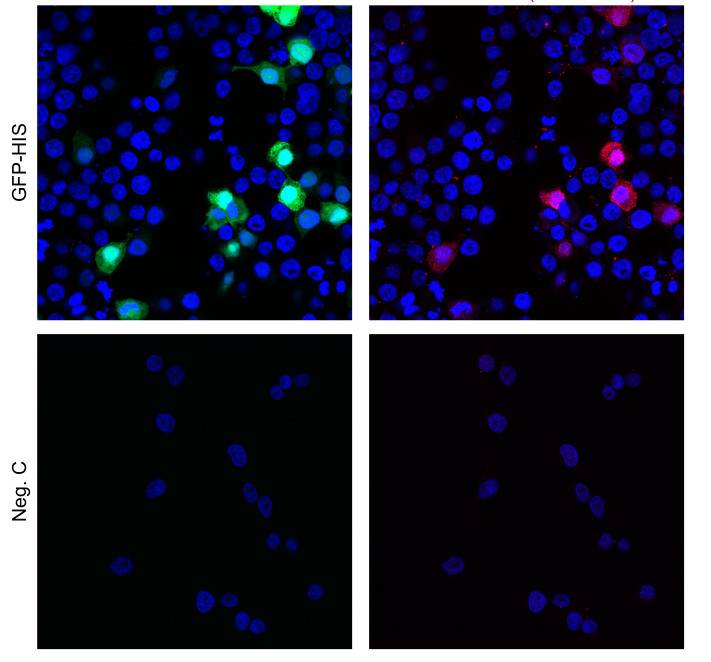
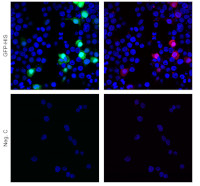
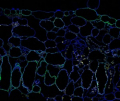
HEK293 cells were transfected with the indicated plasmid (SARS-CoV-2 N myc tagged, PMID: 34799561, TBEV NS3-HA, PMID: 29321318, Rab5 mcherry, Addgene: 4920, GFP-HIS) using genejuice transfection reagent (EMD Millipore) according to the manufacturer’s instructions. After 24 hours of transfection, cells were fixed in 4% formaldehyde and permeabilized in PBS containing 0.5% Triton X-100 and 20 mM glycine. Then, cells were stained with the primary anti-3xHis tag antibodies at a concentration of 1 μg/mL for 1 hour at room temperature. Followed by three washes in PBS. Cells were then stained using secondary antibodies, donkey anti-rabbit Alexa555 (1:500, Thermo Fisher Scientific, a31572) in PBS containing 2% BSA for 1h at RT. Nuclei were stained using DAPI (1 μg/mL). Images were acquired using a Leica SP8 Laser Scanning Confocal Microscope with a 63x oil objective (Leica) and Leica Application Suit X software (LAS X, Leica).
Courtesy of Dr. Anna K Överby, Molecular Infection Medicine Sweden (MIMS),Section of Virology. Department of Clinical Microbiology Umeå University, Sweden - Background
-
Background: His-Tag is a polyhistidine tag which consists of 3 or 6 histidine residues introduced on N- or C-terminus of the protein. The polyhistidine-tag can be used for recombinant protein detection using specific antibodies and it is not conjugated to any dye or enzyme. - Protocols
-
Agrisera Western Blot protocol and video tutorials
Protocols to work with plant and algal protein extracts
Agrisera Educational Poster Collection - Reviews:
-
This product doesn't have any reviews.
Related products

AS09 602 | Clonality: Polyclonal | Host: Goat | Reactivity: Rabbit IgG (H&L)
AS20 4443 | Clonality: Polyclonal | Host: Rabbit | Reactivity: Green Fluorescence Protein (GFP)

AS20 4457 | Clonality: Monoclonal | Host: Mouse | Reactivity: His-tagged proteins