1
Anti-GFP | Green fluorescent protein (VENUS)
AS18 4227 | Clonality: Polyclonal | Host: Rabbit | Reactivity: Venus
- Product Info
-
Immunogen: Full length, recombinat VENUS protein, expressed in E.coli, UniProt: P42212 Host: Rabbit Clonality: Polyclonal Purity: Immunogen affinity purified serum in PBS pH 7.4. Format: Lyophilized Quantity: 50 µg Reconstitution: For reconstitution add 50 µl, of sterile water Storage: Lyophilized antibody can be stored at -20°C for up to 3 years. Once reconstituted make aliquots to avoid repeated freeze-thaw cycles. Please remember to spin the tubes briefly prior to opening them to avoid any losses that might occur from material adhering to the cap or sides of the tube. Tested applications: Western blot (WB) Recommended dilution: 1 : 1000 (WB) Expected | apparent MW: 26 kDa - Reactivity
-
Confirmed reactivity: cell lysate overexoressing Venus protein fusion Not reactive in: No confirmed exceptions from predicted reactivity are currently known - Application Examples
-
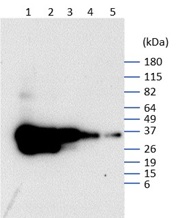
Application example 
200 ng mVenus YFP protein (1); 100 ng mVenus YFP protein (2) ; 49 ng mVenus YFP protein (3) ; 24 ng mVenus YFP protein (4) ; 12 ng mVenus YFP protein (5)
MW markers: BenchMark™ Pre-stained Protein Ladder (10748010)
>0.200 – 0.012 µg of total protein from a pure stock of mVenus YFP protein in 1x SIGMAFAST EDTA-free Protease Inhibitor (S8830-2TAB) and denatured with 1x reducing Laemmli SDS buffer at 90°C for 10 min, and insoluble material pelleted at 20,000 xg for 15 min. Samples were run to separation on a 4-12% SDS-PAGE gel and blotted 7 mins to PVDF using ThermoFisher iBlot high voltage Protocol 3. Blot was blocked with 7.5 % milk in TBS-T for 20mins/RT with agitation. Blot was incubated in the primary antibody (i.e. Rabbit anti-Venus IgG) at a dilution of 1:10 000 with 7.5 % milk in TBS-T ON/4°C with agitation. The antibody solution was decanted and the blot was rinsed briefly three times, then washed once for 15 min and 3 times for 5 min in TBS-T at RT with agitation. Blot was blocked with 7.5 % milk in TBS-T for 20mins/RT. Blot was then incubated in Agrisera matching secondary antibody (anti-rabbit IgG horse radish peroxidase conjugated) diluted to 1:10 000 in TBS-T for 2.5h/RT with agitation. The blot was washed as above, then as above with TBS, and developed for 5 min with chemiluminescent detection reagent, according to manufature's instruction.
Exposure time was approximately 3 seconds.Courtesy of Dr. Dr Nanakow Baiden at Rothamsted Research, UK
Application examples: 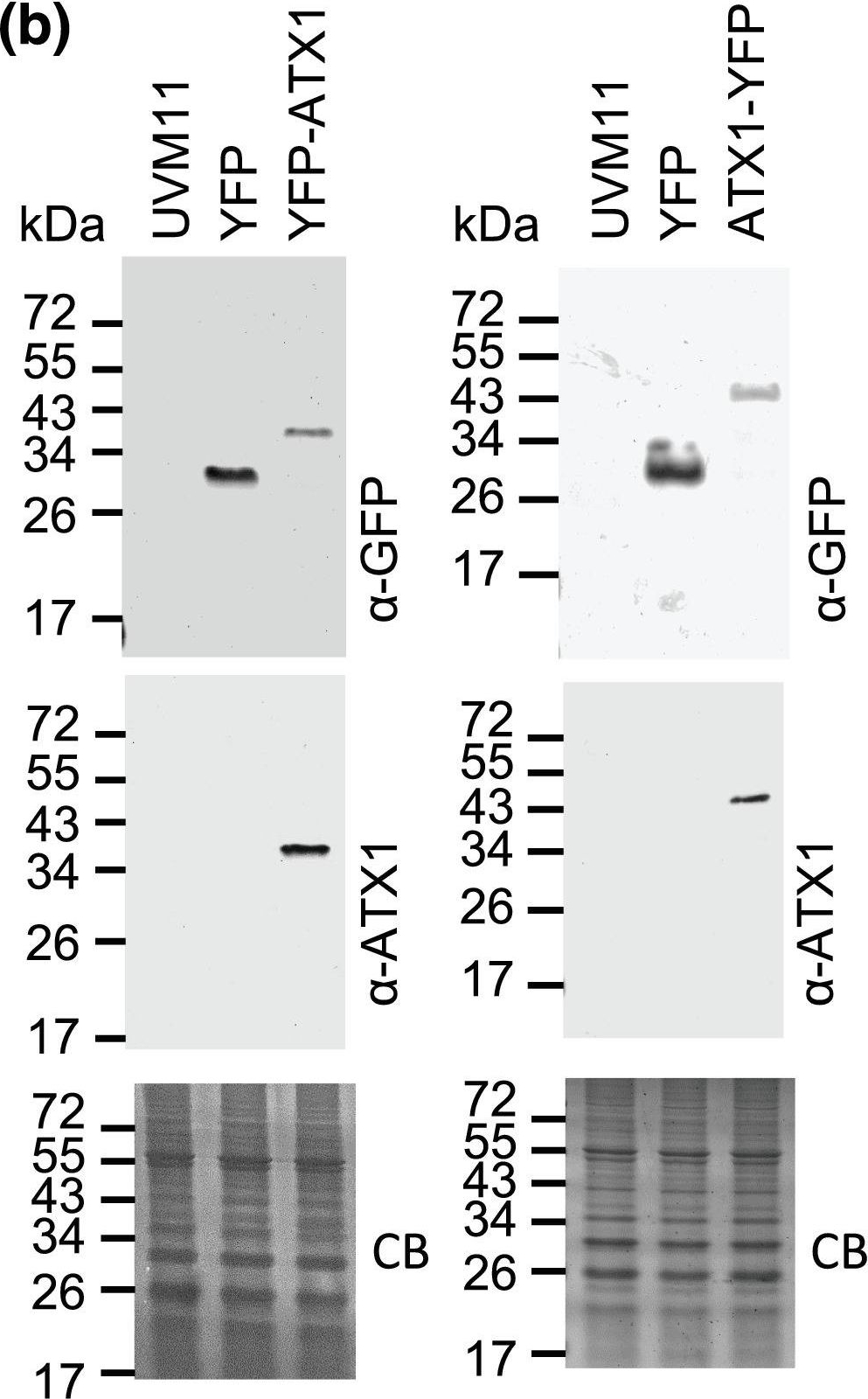
Reactant: Chlamydomonas reinhardtii (Green Alga)
Application: Western Blotting
Pudmed ID: 35141461
Journal: Plant Direct
Figure Number: 3B
Published Date: 2022-02-01
First Author: Pham, K. L. J., Schmollinger, S., et al.
Impact Factor: None
Open PublicationYFP?Atx1 is expressed in Chlamydomonas. (a) Physical map of the YFP control, the YFP?ATX1, and the ATX1?YFP construct. (b) Total protein from UVM11 (parental strain) and UVM11 transformed strains expressing either the N?terminal YFP?ATX1 fusion protein or the C?terminal ATX1?YFP fusion protein as well as YFP were separated by 15% SDS PAGE and after transfer to a nitrocellulose membrane probed using a GFP antibody. ATX1 antibody was also tested in the assay to confirm cross?reactivity with the fusion protein. Coomassie blue stain (CB) of the gel is shown as a loading control. The expected size of YFP is 26.7?kDa, of the N?terminal YFP?ATX1 fusion protein is 34.2?kDa, and of the C?terminal ATX1?YFP fusion protein is 40.1?kDa
- Background
-
Background: Green fluorescent protein (Venus) from Aequorea victoria (Jellyfish), can be mutated to emit at different wavelengths such as blue for BFP (when Tyr-66 is replaced by His), cyan for CFP (when Tyr-66 is replaced by Trp), and yellow for YFP (when THR-203 is replaced by Tyr). It is an energy-transfer acceptor. Its role is to transduce the blue chemiluminescence of the protein aequorin into green fluorescent light by energy transfer. - Product Citations
-
Selected references: Zhang et al. (2024). An epigenetically mediated double negative cascade from EFD to HB21 regulates anther development. Nat Commun. 2024 Sep 6;15(1):7796. doi: 10.1038/s41467-024-52114-x.
Baiden et al. (2022). Heterologous expression of antimicrobial peptides S-thanatin and bovine lactoferricin in the marine diatom Phaeodactylum tricornutum enhances native antimicrobial activity against Gram-negative bacteria, Algal Research, Volume 69, 2023, 102927, ISSN 2211-9264, https://doi.org/10.1016/j.algal.2022.102927. - Protocols
-
Agrisera Western Blot protocol and video tutorials
Protocols to work with plant and algal protein extracts
Agrisera Educational Posters Collection - Reviews:
-
This product doesn't have any reviews.
Related products

AS09 602 | Clonality: Polyclonal | Host: Goat | Reactivity: Rabbit IgG (H&L)

AS15 2998 | Clonality: Polyclonal | Host: Chicken | Reactivity: native and recombinant GFP