Visar bild 1 av
1

Anti-H3T6pK9me1 | Histone H3 methylated at Lys9 (K9)
Product no: AS16 3174
AS16 3174 | Clonality: Polyclonal | Host: Rabbit | Reactivity: Chicken, C.elegans, D. melanogaster, Human, Mouse, plant, Rat, Xenopus sp.
565
€
Customer reviews
Delivery:
3-6 business days
- Product Info
-
Immunogen: KLH-conjugated synthetic peptide Host: Rabbit Clonality: Polyclonal Purity: Immunogen affinity purified serum. Format: Liquid Quantity: 50 µg Storage: Store lyophilized/reconstituted at -20°C; once reconstituted make aliquots to avoid repeated freeze-thaw cycles. Please remember to spin the tubes briefly prior to opening them to avoid any losses that might occur from material adhering to the cap or sides of the tube. Tested applications: Chromatin immunoprecipitation (ChIP), Dot blot (Dot), Immunofluorescence (IF), Immunohistochemistry (IHC), Western blot (WB) Recommended dilution: 2-5 µg/million cells (ChIP), 1 : 1000 (Dot), 1 : 100 (IF), 1 : 200 (IHC), 1 : 500-1 : 1000 (WB) Expected | apparent MW: 15 kDa
- Reactivity
-
Confirmed reactivity: Caenorhabditis elegans, Human Predicted reactivity: Chicken, Drosophila melanogaster, Human, Mouse, Plant, Rat, Xenopus sp. Not reactive in: No confirmed exceptions from predicted reactivity are currently known - Application Examples
-
application example 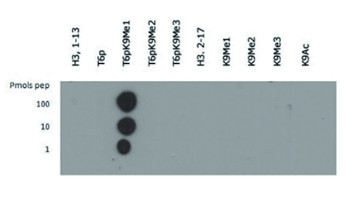
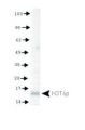
Dot blot using anti-H3T6pK9me1 antibodies. Lane 1: Histone H3 1-13. Lane 2: T6p. Lane 3: T6pK9Me1. Lane 4: T6pK9Me2. Lane 5: T6pK9Me3. Lane 6: Histone H3 2-17. Lane 7: K9Me1. Lane 8: K9Me2. Lane 9: K9Me3. Lane 10: K9Ac. Load: 1, 10, and 100 picomoles of peptide. Primary antibody diluted 1:1 000 for 45 min at 4°C. Secondary antibody: Dylight®488 rabbit secondary antibody at 1:10,000 for 45 min at RT.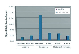
Chromatin Immunoprecipitation using anti-H3T6pK9me1 antibodies. Chromatin from one million formaldehyde cross-linked Hela cells was used with 2ug of H3T6pK9me1 and 20ul of magnetic IgG beads per immunoprecipitation. A no antibody (No Ab) control was also used. Immunoprecipitated DNA was quantified using quantitative real-time PCR and normalized to the input chromatin.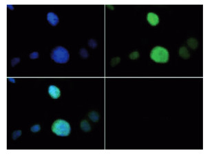
Immunofluorescence using anti-H3T6pK9me1 antibodies. Tissue: HeLa cells. Fixation: 0.5% PFA. Primary antibody: incubated at a 1:100 dilution for 1 h at RT. Secondary antibody: FITC secondary antibody at 1:10 000 for 45 min at RT. Localization: H3T6pK9me1 is nuclear and chromosomal. Staining: H3T6pK9me1 is expressed in green and the nuclei are counterstained with DAPI (blue).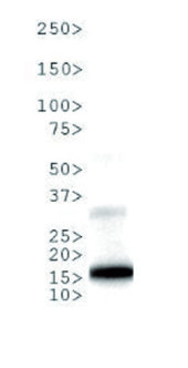
Western Blot using anti-H3T6pK9me1 antibodies. 30 μg NIH-3T3 Histone extracts. Primary antibody diluted 1:1 000 overnight at 4°C. Secondary antibody: IRDye800™ rabbit secondary antibody at 1:10 000 for 45 min at RT.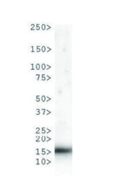
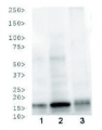
Western Blot anti-H3T6pK9me1 antibodies. 30 μg C. elegans embryo lysate. Primary antibody diluted 1:1 000 overnight at 4°C. Secondary antibody: IRDye800™ rabbit secondary antibody at 1:10 000 for 45 min at RT.
Western Blot using anti-H3T6pK9me1 antibodies. 30 μg HeLa Histone extracts. Primary antibody diluted 1:1 000 overnight at 4°C. Secondary antibody: IRDye800™ rabbit secondary antibody at 1:10 000 for 45 min at RT. - Additional Information
-
Additional information: This antibody preparation is provided in 20 mM Potassium Phosphate pH 7,2, 150 mM NaCl, 0,01% sodium azide and 30% glycerol - Background
-
Background: Methylation of Histone H3 at Lys9 (K9) is an epigenetic silencer of transcription. Gene silencing from histone post translational modifications, as well as DNA methylation, play a key role in the development of normal tissues. - Reviews:
-
This product doesn't have any reviews.
Related products

AS09 602 | Clonality: Polyclonal | Host: Goat | Reactivity: Rabbit IgG (H&L)
219 €
AS16 3173 | Clonality: Polyclonal | Host: Rabbit | Reactivity: C.elegans, D. melanogaster, Human, Mouse, plant, Rat, Xenopus sp.
518 €
AS16 3170 | Clonality: Polyclonal | Host: Rabbit | Reactivity: C.elegans, D. melanogaster, Human, Mouse, plant, Rat, Xenopus sp.
565 €