Visar bild 1 av
1
Anti-H3T3pK4ac | Histone H3 (ac Lys4, p Thr3)
Product no: AS16 3170
AS16 3170 | Clonality: Polyclonal | Host: Rabbit | Reactivity: C.elegans, D. melanogaster, Human, Mouse, plant, Rat, Xenopus sp.
565
€
Customer reviews
Delivery:
3-6 business days
- Product Info
-
Immunogen: KLH-conjugated synthetic acetylated/phosphorylated peptide surrounding Lysine 4 and Threonine 3 of human Histone H3 Host: Rabbit Clonality: Polyclonal Purity: Immunogen affinity purified serum. Format: Liquid Quantity: 50 µg Storage: Store lyophilized/reconstituted at -20°C; once reconstituted make aliquots to avoid repeated freeze-thaw cycles. Please remember to spin the tubes briefly prior to opening them to avoid any losses that might occur from material adhering to the cap or sides of the tube. Tested applications: Chromatin immunoprecipitation (ChIP), Dot blot (Dot), Immunofluorescence (IF), Immunohistochemistry (IHC), Western blot (WB) Recommended dilution: 2-5 µg/million cells (ChIP), 1 : 1000 (Dot), 1 : 100 (IF), 1 : 50 (IHC), 1 : 500 (WB) Expected | apparent MW: 15 kDa
- Reactivity
-
Confirmed reactivity: Caenorhabditis elegans, Human Predicted reactivity: Chicken, Drosophila melanogaster, Mouse, Plant, Rat, Xenopus sp. Not reactive in: No confirmed exceptions from predicted reactivity are currently known - Application Examples
-
application example 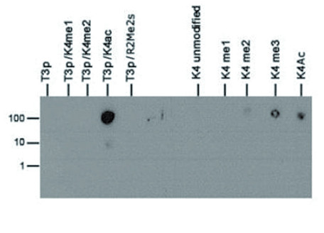
Dot Blot using anti-H3T3pK4ac antibodies. Lane 1: T3p. Lane 2: T3pK4me1. Lane 3: T3pK4me2. Lane 4: T3pK4ac. Lane 5: T3pR2me2s. Lane 6: K4 unmodified. Lane 7: K4me1. Lane 8: K4me2. Lane 9: K4me3. Lane 10: K4ac. Load: 1, 10, and 100 picomoles of peptide. Primary antibody used at 1:1,000 dilution for 45 min at 4°C. Secondary antibody: Dylight®488 rabbit secondary antibody at 1:10 000 for 45 min at RT.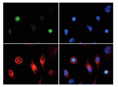
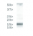
Immunofluorescence using anti-H3T3pK4ac antibodies. Tissue: HeLa cells. Fixation: 0.5% PFA. Primary antibody used at a 1:100 dilution for 1 h at RT. Secondary antibody: Dylight® 488 secondary antibody at 1:10 000 for 45 min at RT. Localization: Histone H3T3pK4ac is nuclear and chromosomal. Staining: H3T3pK4ac is expressed in green while the nuclei and aplpha-tubulin were coexpressed with DAPI (blue) and Dylight® 550 (red).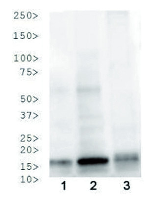
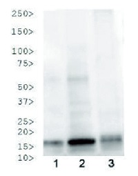
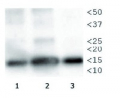
Western Blot using anti-phospho-acetyl-Histone H3 antibodies (H3T3pK4ac). Lane 1: HeLa histone extracts. Lane 2. NIH-3T3 histone extracts. Lane 3: C. elegans embryo lysate. Load: 30 μg per lane. Primary antibody used at 1:500 overnight at 4°C. Secondary antibody: IRDye800™ rabbit secondary antibody at 1:10 00 for 45 min at RT. - Additional Information
-
Additional information: This antibody preparation is provided in 20 mM Potassium Phosphate pH 7,2, 150 mM NaCl, 0,01% sodium azide and 30% glycerol - Background
-
Background: Chromatin is the arrangement of DNA and proteins in which chromosomes are formed. Correspondingly, chromatin is formed from nucleosomes, which are comprised of a set of four histone proteins (H2A, H2B, H3, H4) wrapped with DNA. Chromatin is a very dynamic structure in which numerous post-translational modifications work together to activate or repress the availability of DNA to be copied, transcribed, or repaired. These marks decide which DNA will be open and commonly active (euchromatin) or tightly wound to prevent access and activation (heterochromatin). Common histone modifications include methylation of lysine and arginine, acetylation of lysine, phosphorylation of threonine and serine, and sumoylation, biotinylation, and ubiquitylation of lysine. Phosphorylation of threonine 3 (H3T3p) is a known mitotic marker and modified by the Haspin/Thr3 enzyme, while acetylation of lysine 4 (H3K4ac) on histone 3 is associated with transcriptional activation by Esa1.
Alternative names: H3.3B, H3 histone, family 3A, H3.3AH3F3H3F3B, histone H3.3, MGC87782, MGC87783, H3pT3/K4ac . - Reviews:
-
This product doesn't have any reviews.
Related products

AS09 602 | Clonality: Polyclonal | Host: Goat | Reactivity: Rabbit IgG (H&L)
219 €
AS16 3169 | Clonality: Polyclonal | Host: Rabbit | Reactivity: C.elegans, D. melanogaster, Human, Mouse, plant, Rat, Xenopus sp.
565 €
AS16 3168 | Clonality: Polyclonal | Host: Rabbit | Reactivity: C.elegans, D. melanogaster, Human, Mouse, plant, Rat, Xenopus sp.
565 €