1
Anti-HSP16,9 | 16,9 kDa class I heat shock protein 2
AS12 2570 | Clonality: Polyclonal | Host: Rabbit | Reactivity: Triticum aestivum
- Product Info
-
Immunogen: Recombinant protein of Triticum aestivum UniProt: Q41560 Host: Rabbit Clonality: Polyclonal Purity: Serum Format: Lyophilized Quantity: 100 µl Reconstitution: For reconstitution add 100 µl of sterile water Storage: Store lyophilized/reconstituted at -20°C; once reconstituted make aliquots to avoid repeated freeze-thaw cycles. Please remember to spin the tubes briefly prior to opening them to avoid any losses that might occur from material adhering to the cap or sides of the tube. Tested applications: Western blot (WB) Recommended dilution: 1 : 1000-1 : 10 000 (WB) Expected | apparent MW: 16.8 kDa - Reactivity
-
Confirmed reactivity: Triticum aestivum Predicted reactivity: Aegilops kotschyi, Hordeum vulgare, Triticum sp.
Species of your interest not listed? Contact usNot reactive in: No confirmed exceptions from predicted reactivity are currently known - Application Examples
-
Application example 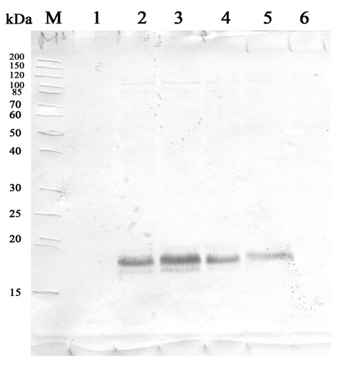
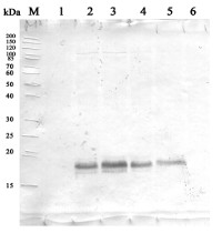
24 µg of Triticum aestivum L. whole leaf extract (1), 23 µg of Triticum aestivum L. whole leaf extract 37°С,3h (2), 22 µg of Triticum aestivum L. whole leaf extract, 37°С, 24h (3), 20 µg of Triticum aestivum L. whole leaf extract, 37°С,24h+50°C/1h (4), 17 µg of Triticum aestivum L. whole leaf extract, 37°С,24h+50°C, 3h (5), 23 µg of Triticum aestivum L. whole leaf extract, Control+50°C, 1h (6).
700 µg of total protein from spring wheat Triticum aestivum L. green leaves extracted with write exact buffer components 100 mM Tris HCl (pH=7.4), 1 mM beta-mercaptoethanol, 1 mM PMSF and denatured with 65.2 mM Tris HCl (pH=6.8), 1mM EDTA, 1% SDS, 20% glycerol, 5% beta-mercaptoethanol at 97°C for 5 min and 20 μg of total protein were separated on 12.5 % SDS-PAGE and blotted 2h on nitrocellulose membrane (GE Healthcare) using tank transfer. Blots were blocked with a skimmed milk 3 % in TBS for 1h at room temperature (RT) with agitation. Blot was incubated in the primary antibody at a dilution of 1: 1 000 for 1h at RT with agitation in TBS. The antibody solution was decanted and the blot washed 2 times for 10 min in TBS-T at RT with agitation. Blot was incubated in secondary antibody (anti-rabbit IgG, ALP conjugated, AS09 607, from Agrisera) diluted to 1:1000 in a skimmed milk 3 % in TBS for 1h at RT with agitation. The blot was washed 3 times for 5 min in TBS-T at RT with agitation and developed WB. The proteins were detected with 5-bromo-4-chloro-3-indolyl phosphate and Nitrotetrazolium Blue. Exposure time was 5.20 minutes.
Courtesy of Dr. Olga Borovik, Laboratory of Physiological Genetics Siberian Institute of Plant Physiology and Biochemistry SB RAS Russia

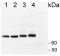
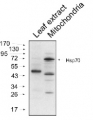
From 0.1-200 ng of recombinant HSP16B protein from Triticum aestivum were separated on 15 % SDS-PAGE and blotted 1h to nitrocellulose using semi-dry transfer at 15 V for 1 hour in a buffer containing: 48mM Tris, 39 mM Glycine, 0.02% SDS, 20 % methanol, pH 9.2. Blots were blocked with with 2.5% milk in TBS (50mM Tris, 150 mM NaCl, pH 7.5) for 1 hour at room temperature (RT) with agitation. Blot was incubated in the primary antibody at a dilution of 1: 10 000 for 2h at RT TBS with 2.5% milk with agitation. Three washes (15 minutes each) were carried out with TBS and 0.05% Tween20 at RT with agitation. Blot was incubated in secondary antibody (anti-rabbit IgG horse radish peroxidase conjugated, diluted to 1:10 000 in for 1h at RT with agitation. The blot was washed as above and developed using Pierce western blotting substrate (#32106). The exposure time used for visualization was 6.5 minutes.
Courtesy of Indu Santhanagopalan, Dept. of Biochem and Mol Biology, Lederle Graduate Research Center, USA.
- Background
-
Background: Hsp16.9 belongs to a family of class I of a small heat shock proteins (HSP20) family. Localized in cytoplasm. Alternative protein name: Heat shock protein 16.9B
- Product Citations
-
Selected references: Fedotova et al. (2020). Influence of high temperatures on heat tolerance and synthesis of heat shock proteins in spring wheat at the initial stages of development // Siberian Journal of Life Sciences and Agriculture. 2020. ?. 12, ? 5. C. 179-191. DOI: 10.12731/2658-6649-2020-12-5-179-191 - Reviews:
-
This product doesn't have any reviews.
Related products

AS09 602 | Clonality: Polyclonal | Host: Goat | Reactivity: Rabbit IgG (H&L)
AS08 371 | Clonality: Polyclonal | Host: Rabbit | Reactivity: A. thaliana, B. juncea, B. oleracea, C. sativus, C. reinhardtii, D. subspicatus, E. tef, G. vermiculophylla, H. vulgare, M. sativa, O. sativa, P. kurroa, P. strobus, Salicornia sp., S. italica. S. vulgaris, S. lycopersicum, Trebouxia TR1 and TR9, T. aestivum, Triticum turgidum ssp. durum, Z. mays, P. falciparum, V. faba
Benefits of using this antibody
AS08 347 | Clonality: Polyclonal | Host: Rabbit | Reactivity: Arabidopsis thalian, Brassica oleracea, Hordeum vulagre