1
Anti-LHCR4 | Protein fucoxanthin chlorophyll a/c protein
AS19 4303 | Clonality: Polyclonal | Host: Rabbit | Reactivity: Phaeodactylum tricornutum
- Product Info
-
Immunogen: KLH-conjugated peptide derived from Phaeodactylum tricornutum LHCR4, UniProt: B7FQE1 Host: Rabbit Clonality: Polyclonal Purity: Immunogen affinity purified serum in PBS pH 7.4. Format: Lyophilized Quantity: 50 µg Reconstitution: For reconstitution add 50 µl, of sterile water Storage: Store lyophilized/reconstituted at -20°C; once reconstituted make aliquots to avoid repeated freeze-thaw cycles. Please remember to spin the tubes briefly prior to opening them to avoid any losses that might occur from material adhering to the cap or sides of the tube. Tested applications: Western blot (WB) Recommended dilution: 1 : 1000 (WB) Expected | apparent MW: 23 kDa - Reactivity
-
Confirmed reactivity: Phaeodactylum tricornutum Predicted reactivity: Gambierdiscus australes, Madurella mycetomatis
Species of your interest not listed? Contact us
Not reactive in: No confirmed exceptions from predicted reactivity are currently known - Application Examples
-
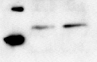
Samples: 1 - 25 µg of Phaeodactylum tricornutum whole cell extract from low light (35 µmol photons m-2 s-1) acclimated WT cells 2 – 25 µg of Phaeodactylum tricornutum whole cell extract from low light (35 µmol photons m-2 s-1) acclimated WT cells exposed to high light (1000 µmol photons m-2 s-1) for 1 hour.
Total protein was extracted from P. tricornutum cell pellets that had been flash-frozen in liquid nitrogen and stored in -80 °C with a buffer containing 2% SDS, 6M Urea and 50 mM Tris, pH 6.8. A five mm pre-cooled (-80°C) stainless steel bead (QIAGEN) were added to each of the tubes with frozen cell pellets, and the cells were mechanically broken and homogenized in two steps using the TissueLyser system (QIAGEN). The samples were first placed in a precooled (-80°C) adapter set followed by cell disruption for 2 min at 25 Hz. Before the second shaking step (8 min at 25 Hz), the samples were transferred to a room temperature adapter set and 700 µl lysis buffer (2% SDS, 6M Urea and 50 mM Tris, pH 6.8) were added. Insoluble material was removed by centrifugation (100 G) for 30 min at 4°C following the protocol from Juhas et al. (2014). The lysate was stored at -80°C. 25 µg of WT samples were loaded on the gel. A loading dye containing DTT, SDS, glycin, Tris (pH 6.8) and BPB was added to the samples and the samples were heated at 40°C for 10 min. Proteins were separated on 12 % SDS-PAGE and blotted 1h at 100V to nitrocellulose (pore size of 0.2 µm), using wet transfer. Blot was blocked with 5 % milk for 1h/RT with agitation. Blot was incubated in the primary antibody at a dilution of 1: 1 000 ON/4°C with agitation in 5% milk in PBS-T. The antibody solution was decanted, and the blot was washed 4 times for 5 min in PBS-T at RT with agitation. Blot was incubated in goat anti-Rabbit IgG secondary antibody conjugated to HRP (Agrisera, AS09 602l) diluted to 1:25 000 in 5% milk in PBS-T for 2h/RT with agitation. The blot was washed as above before adding AgriseraECL SuperBright chemiluminescent substrate with extreme low femtogram detection range, to the blot. 10 minutes after adding the chemiluminescent substrate, a G:BOX ChemiXRQ gel doc system (Syngene) was used to take pictures of the blots. The exposure time was automatically estimated by the G:BOX software to around 2 minutes. LHCR4 antibody is producing a band of correct size can be seen at maximum contrast for the WT sample in addition to the unspecific bands.
Courtesy od Dr. Marianne Nymark, Institute of Biology,NTNU, Trondheim, Norway - Background
-
Background: LHCR4 (Protein fucoxanthin chlorophyll a/c protein) is a PSI supercomplex peripheral antenna protein, identified by proteome analysis as type‐R light‐harvesting complexes (LHCr4). - Protocols
-
Agrisera Western Blot protocol and video tutorials
Protocols to work with plant and algal protein extracts
Agrisera Educational Poster Collection - Reviews:
-
This product doesn't have any reviews.
Related products

AS09 602 | Clonality: Polyclonal | Host: Goat | Reactivity: Rabbit IgG (H&L)
AS05 084 | Clonality: Polyclonal | Host: Rabbit | Reactivity: [global antibody] for higher plants, algae, liverwort, cyanobacteria, diatoms | cellular [compartment marker] of thylakoid membrane
Benefits of using this antibody
AS03 037 | Clonality: Polyclonal | Host: Rabbit | Reactivity: global antibody and compartment marker for higher plants, lichens, algae, cyanobacteria, dinoflagellates, diatoms
Benefits of using this antibody