1
Anti-mNeonGreen | Fluorescent Protein
AS21 4525 | Clonality: Polyclonal | Host: Rabbit | Reactivity: mNeonGreen | Fluorescent Protein
- Product Info
-
Immunogen: KLH-conjugated peptide derived from synthetic peptide, UniProt: A0A1S4NYF2 from common lancelet Branchiostoma lanceolatum Host: Rabbit Clonality: Polyclonal Purity: Immunogen affinity purified serum, in PBS pH 7.4. Format: Lyophilized Quantity: 50 µg Reconstitution: For reconstitution add 50 µl, of sterile water. Storage: Store lyophilized/reconstituted at -20°C; once reconstituted make aliquots to avoid repeated freeze-thaw cycles. Please remember to spin the tubes briefly prior to opening them to avoid any losses that might occur from material adhering to the cap or sides of the tube. Tested applications: Immunofluorescence (IF), Western blot (WB) Recommended dilution: 1 µg/1ml (IF), 1 : 1000 - 1: 5000 (WB) Expected | apparent MW: Depends upon used fusion partner - Reactivity
-
Confirmed reactivity: mNG tag in plant (Arabidopsis thaliana) and algal vectors (Chlamydomonas reinhardtii) Predicted reactivity: Species of your interest not listed? Contact us Not reactive in: No confirmed exceptions from predicted reactivity are currently known. - Application Examples
-
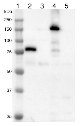
Samples:
1- Precision Plus Protein Standards (Bio-Rad)
2-Arabidopsis thaliana expressing mNeonGreen-tagged fusion protein
3-Arabidopsis thaliana wildtype total cell extract
4-Chlamydomonas reinhardtii expressing mNeonGreen-tagged fusion protein
5-Chlamydomonas reinhardtii wildtype cell extract
20 µg of total protein extracted from Arabidopsis thaliana leaves or Chlamydomonas reinhardtii cells with 62.5 mM Tris-HCl pH 6.8, 10 % glycerol, 2 % SDS, 50 mM DTT, 1x protease inhibitors (P9599, Sigma) and heated at 65ºC for 5 minutes were separated on 10 % SDS-PAGE and blotted 1 h to PVDF membrane (pore size 0.45 µm) using wet transfer. The blot was blocked with 5 % milk/TBS-T for 1 h/RT with agitation and then incubated in the primary antibody at a dilution 1: 1000 in 5 % milk/TBS-T for 1h/RT with agitation. The antibody solution was decanted, and the blot was briefly rinsed, then washed three times for 10 minutes in TBS-T with agitation. The blot was incubated in Agrisera matching secondary antibody (anti-rabbit IgG, HRP conjugated, AS09 602) at a dilution of 1: 25 000 in 5 % milk/TBS-T for 1h/RT with agitation. The blot was washed as above, developed for 2 minutes with AgriseraECLSuperBright substrate and imaged with a Bio-Rad ChemiDoc MP digital imaging system. Exposure time was 2.5 s.
Courtesy of Hanie Khorshidi, University of Saskatchewan, Canada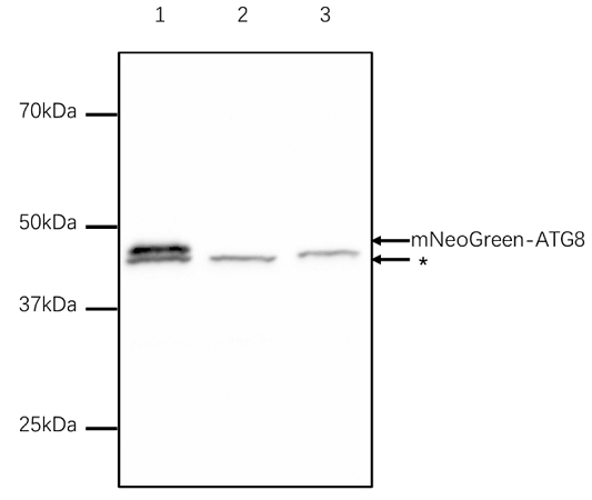
Samples:
1 - Nicotiana benthamiana whole leaf extract expressing mNeonGreen-ATG8
2 – Nicotiana benthamiana whole leaf extract expressing YFP-ATG8
3 – Nicotiana benthamiana whole leaf extract expressing mCherry-ATG8
MW markers: (Biorad)Precision Plus Protein Dual Color Standards, 500 µl #161037450 µg of total protein extracted freshly from Nicotiana benthamiana leaves with exaction buffer (50mM Tris-HCl pH7.5, 150mM NaCl, 10%(v/v) glycerol, 0.5mM EDTA and 0.5% NP40) denatured with SDS-sample buffer at 95°C for 5 min, proteins were separated on 10 % SDS-PAGE and blotted 0.5h to PVDF membrane using semi-dry transfer. Blots were blocked with 5 % milk in PBS-T for 1h/RT with agitation. Blot was incubated in the primary antibody at a dilution of 1: 3 000 for ON/4°C with agitation. The antibody solution was decanted, and the blot was rinsed briefly twice, then washed once for 15 min and 3 times for 5 min in PBS-T at RT with agitation. Blot was incubated in Agrisera matching secondary antibody (anti-rabbit IgG (H&L), horse radish peroxidase conjugated, AS09 602) diluted to 1: 5 000 in for 1h/RT with agitation. The blot was washed as above, developed for 5 mins with Agrisera ECL Bright and detected with film.
Courtesy of Dr. Jiaqi Sun, McGill University, Canada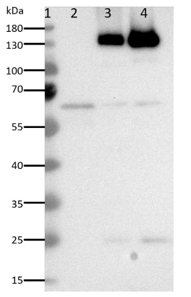
Samples:
1- PageRuler™ Prestained Protein Ladder (Thermo Fisher)
2- 20 µg Arabidopsis thaliana wildtype microsomal membrane preparation
3- 20 µg Arabidopsis thaliana expressing VHA-a1-GSL-NeonGreen microsomal membrane preparation
4- 20 µg Arabidopsis thaliana expressing VHA-a3-GSL-NeonGreen microsomal membrane preparation20 µg of microsomal membrane proteins were prepared from Arabidopsis thaliana 4-week -old rosette leaves with a buffer containing 350mM sucrose, 70mM Tris-HCl pH8, 10% glycerol, 3mM Na2EDTA, 0.15% BSA, 1.5% PVP-40, 4mM DTT, 1x Roche completeTM protease Inhibitor and denatured at 95ºC for 5 minutes. Proteins were separated on 10 % SDS-PAGE and blotted for 1 h to PVDF membrane (pore size 0.2 µm) using wet transfer. The blot was blocked with 5% milk/TBS-T (0.05%) for 1 h at RT with agitation and then incubated in the primary antibody at a dilution 1: 1000 in 5% milk/TBS-T (0.05%) for 1h at RT with agitation. The antibody solution was decanted, and the blot was briefly rinsed, then washed three times for 10 minutes in TBS-T (0.05%) with agitation. The blot was incubated in Agrisera matching secondary antibody (anti-rabbit IgG, HRP conjugated, AS09 602) at a dilution of 1: 25 000 in 5 % milk/TBS-T (0.05%) for 1hr at RT with agitation. The blot was washed as above, developed for 1 min with AgriseraECLBright substrate and imaged with a cooled CCD camera system (Intas ADVANCED Fluoreszenz u. ECL Imager). Exposure time was 10s.
Courtesy of Upendo Lupanga, Centre for Organismal Studies, University of Heidelberg, Germany
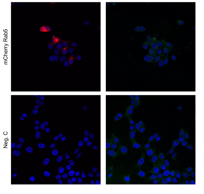
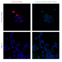
HEK293 cells were transfected with the indicated plasmid (SARS-CoV-2 N myc tagged, PMID: 34799561, TBEV NS3-HA, PMID: 29321318, Rab5 mcherry, Addgene: 4920, GFP-HIS) using genejuice transfection reagent (EMD Millipore) according to the manufacturer’s instructions. After 24 hours of transfection, cells were fixed in 4% formaldehyde and permeabilized in PBS containing 0.5% Triton X-100 and 20 mM glycine. Then, cells were stained with the primary anti-mNeonGreen epitope tag, rabbit polyclonal antibodies at a concentration of 1 μg/mL for 1 hour at room temperature. Followed by three washes in PBS. Cells were then stained using secondary antibodies, donkey anti-rabbit Alexa488 (1:500, Thermo Fisher Scientific, a21206 in PBS containing 2% BSA for 1h at RT. Nuclei were stained using DAPI (1 μg/mL). Images were acquired using a Leica SP8 Laser Scanning Confocal Microscope with a 63x oil objective (Leica) and Leica Application Suit X software (LAS X, Leica). Anti-NS3 antibodies are used as a control. Left panel anti-mCheery rab6, right panel protein with mCherry construct, recognized by mNeonGreen antibodies.
Courtesy of Dr. Anna K Överby, Molecular Infection Medicine Sweden (MIMS),Section of Virology. Department of Clinical Microbiology Umeå University, Sweden
- Additional Information
-
Additional information: The peptide used to elicit this antibody is conserved in the following expression vectors:
- dCas9-P2A-DHFR(TSc3)-T2A-mNeonGreen [Cloning vector pBM020]
- Cas9-P2A-DHFR(TSc3)-T2A-mNeonGreen [Cloning vector pBM006]
- mNeonGreen4-tDeg [Cloning vector pminiCMV-mNeonGreen4-tDeg]
- ER-localized mNEONGREEN [Binary vector pKT-NM-erNEON]
- mNeonGreen-3xFLAG [Cloning vector pLM160-mNeonGreen]
- mNeonGreen-ty1 [Cloning vector pSAG1-mNeonGreen_TUB1-dTomato]
- mNeonGreen, partial [Binary vector pRATIO2131]
The peptide used to elicit this antibody is not conserved in GFP protein sequence. Antibody is also recognzing mCheery sequence.Additional information (application): This antibody is detecting protein sequence of mNG tag in plant and algal vectors.
This antibody is also reacting to some degree with YFP and mCherry. - Background
-
Background: mNeonGreen (Fluorescent Protein) is a new bright monomertic yellow-green fluorescent, with low conservation level to GFP. It has an excitation maximum at 506 nm and an emission maximum at 517 nm and therefore is compatible with the most GFP filter sets. mNeonGreen is 3x brighter compare to GFP, more stable and does not bleach so fast as GFP, which makes it very suitable for confocal and super resolution microscopy, for fusion proteins with low expression levels. It can be used in bicistronic vectors, which will allow simultaneous expression of two proteins separately, from the same RNA transcript. mNeonGreen has MW of 26.6 kDa. - Protocols
-
Agrisera Western Blot protocol and video tutorials
Protocols to work with plant and algal protein extracts
- Reviews:
-
Wenbin Du | 2024-02-20In our study, we analyzed total protein samples from Arabidopsis leaves tagged with mNeonGreen, using this antibody for Western Blot analysis. We tested antibody dilutions of 1:5,000, and found that the 1:5,000 dilution provided sufficient signal intensity overnight for our fusion protein. Importantly, clear signals were identified at around 105 kDa, matching our fusion protein's expected size. The antibody displayed strong performance in Arabidopsis samples, indicating its effectiveness for future experimental studies. Based on our findings, we recommend continuing to use this antibody and would certainly consider purchasing it again.Katharina König | 2023-10-17We analyzed whole cell samples of a Chlamydomonas reinhardtii chloroplastidic protein tagged with mNeonGreen using 2.5 µg, 5 µg and 10 µg total protein per lane. After Western Blot transfer, we tested antibody dilutions of 1:10.000, 1: 5.000 and 1:3.000, with a dilution of 1:10.000 overnight giving sufficient signal for our over-expressed fusion protein. We obtained signals at approximately 55 kDa for our fusion protein. The antibody worked well for Chlamydomonas reinhardtii and we would buy it again.
Related products

AS09 602 | Clonality: Polyclonal | Host: Goat | Reactivity: Rabbit IgG (H&L)

AS18 4178 | Clonality: Polyclonal | Host: Chicken | Reactivity: mCherry