1
Anti-NBR1 | Autophagy substrate NBR1
AS14 2805 | Clonality: Polyclonal | Host: Rabbit | Reactivity: Arabidopsis thaliana, Physcomitrella patens
- Product Info
-
Immunogen: Host: Rabbit Clonality: Polyclonal Purity: Serum Format: Lyophilized Quantity: 50 µl Reconstitution: For reconstitution add 50 µl of sterile water Storage: Store lyophilized/reconstituted at -20°C; once reconstituted make aliquots to avoid repeated freeze-thaw cycles. Please remember to spin the tubes briefly prior to opening them to avoid any losses that might occur from material adhering to the cap or sides of the tube. Tested applications: Immunolocalization (IL), Western blot (WB) Recommended dilution: 1: 1000 (IL), 1 : 500-1 : 5000 (WB) Expected | apparent MW: 75 | 100 kDa
- Reactivity
-
Confirmed reactivity: Arabidopsis thaliana, Physcomitrium patens Predicted reactivity: Brassicaceae family, Zea mays
Species of your interest not listed? Contact usNot reactive in: Citrus sinensis, Nicotiana tabacum - Application Examples
-
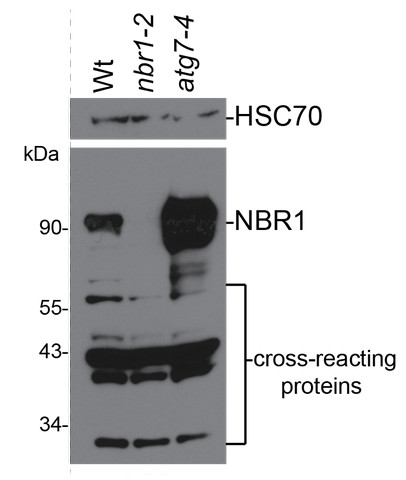
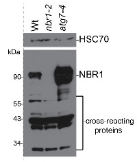
Total protein from approximately eight 8-d-old seedlings of Arabidopsis thaliana was extracted with NuPAGE sample buffer (106 mM Tris-HCl pH 8.5, 141 mM Tris Base, 2% lithium dodecyl sulfate, 0.51 mM EDTA pH 8.0, 10% glycerol, 0.22 mM Coomassie Blue G250, 0.166 mM Phenol Red) with 50 mM dithiothreitol and denatured at 100°C for 5 min. Proteins were separated by electrophoresis on a Bolt 10% Bis-Tris Plus gel (Invitrogen) and transferred for 40 min at 24 V to an Amersham Protran 0.45 µm nitrocellulose membrane (GE Healthcare Life Sciences) using semidry transfer. The membrane was blocked with 8% milk in TBS-T for 2 h at 4°C with agitation. The membrane was incubated in the primary antibody at a dilution of 1:4000 overnight at 4°C with agitation in 8% milk in TBS-T. The antibody solution was decanted and the blot was rinsed briefly twice, then washed 3 times for 5 min in 8% milk in TBS-T at 4°C with agitation. The membrane was incubated in secondary antibody (horse radish peroxidase conjugated goat anti-rabbit IgG) diluted to 1:5000 in 8% milk in TBS-T for 4 h at 4°C with agitation. The membrane was washed 3 times for 5 min in TBS-T at 4°C with agitation and incubated for 2 min with WesternBright chemiluminescent detection reagent (Advansta). Exposure time was 30 seconds.Courtesy of Pierce Young, Rice University, USAApplication examples: 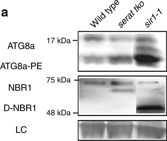
Reactant: Arabidopsis thaliana (Thale cress)
Application: Western Blotting
Pudmed ID: 29079776
Journal: Nat Commun
Figure Number: 5A
Published Date: 2017-10-27
First Author: Dong, Y., Silbermann, M., et al.
Impact Factor: 13.783
Open PublicationAutophagy is specifically induced by limited S-precursor supply for cysteine biosynthesis. a Autophagy induction in the shoot of serat tko and sir1-1 was determined by immunological detection of the canonical autophagy marker ATG8a and NBR1 with specific antisera. Lipidation of ATG8a (ATG8a-PE) (apparent size: 15–20?kDa) is essential for autophagosome formation and indicated by a significant shift during electrophoresis. NBR1 is a cargo receptor for selective autophagy and consequently degraded in autophagic bodies (D-NBR1) (apparent size: 50–75?kDa). b Level of ATG8a-PE and D-NBR1 shown in a were quantified (n?=?3, mean?±?s.e.m., one-way ANOVA, *p?<?0.05). c Autophagy induction in the root of serat tko and sir1-1 was detected by MDC staining. White arrows marked the visible autophagic bodies. Scale bar, 25?µm. d Autophagy induction in the shoot of WT under sulfur deficiency was determined by ATG8a-PE level (n?=?3, mean?±?s.e.m., t-test, *p?<?0.05)
- Additional Information
-
Additional information (application): Specific extraction method and tissue type needs to be used as described in Minina et al, (2013), Dilution in western blot depends upon amount of NBR1 in the sample - Background
-
Background: NBR1 (Autophagy substrate NBR1) is involved in selective autophagy process of damaged organelles, intracellular microbes, protein aggregates, cellular structures and specific soluble proteins. NBR1 mediates the process as an autophagic adapter. The protein has two UBA domains but only the C-terminal UBA domain bound ubiquitin. Alternative names: At4g24690, Putative uncharacterized protein F22K18.110. - Product Citations
-
Selected references: González-Fuente et al. (2026). Bacteria use P-body condensates to attenuate host translation during infection. Sci Adv . 2026 Apr 24;12(17):eaec4477. doi: 10.1126/sciadv.aec4477.
Guan et al. (2025).Arabidopsis phospholipase Dζ2 facilitates vacuolar acidification and autophagy under phosphorus starvation by interacting with VATD. Cell Rep. 2025 Jul 15;44(7):116024. doi: 10.1016/j.celrep.2025.116024.
Hoffman-Sommer et al. (2025). The TRAPPC8/TRS85 subunit of the Arabidopsis TRAPPIII tethering complex regulates endoplasmic reticulum function and autophagy. Plant Physiol. 2025 Mar 1;197(3):kiaf042. doi: 10.1093/plphys/kiaf042.
Julian et al. (2025). ATG8ylation of vacuolar membrane protects plants against cell wall damage. Nat Plants. 2025 Feb;11(2):321-339.doi: 10.1038/s41477-025-01907-z.
Yan et al. (2024). Autophagy modulates Arabidopsis male gametophyte fertility and controls actin organization. Nat Commun . 2024 Nov 21;15(1):10071. doi: 10.1038/s41467-024-54468-8.
Mosesso et al. (2024). Arabidopsis CaLB1 undergoes phase separation with the ESCRT protein ALIX and modulates autophagosome maturation. Nat Commun. 2024 Jun 19;15(1):5188. doi: 10.1038/s41467-024-49485-6.
Lan et al. (2024).Clathrin Light Chains negatively regulate plant immunity by hijacking the autophagy pathway. Plant Commun. 2024 Apr 30:100937.doi: 10.1016/j.xplc.2024.100937.
Zeng et al. (2023) The plant unique ESCRT component FREE1 regulates autophagosome closure. Nat Commun. 2023 Mar 30;14(1):1768. doi: 10.1038/s41467-023-37185-6.
Rodriguez et al. (2020). Autophagy mediates temporary reprogramming and dedifferentiation in plant somatic cells. bioRxiv doi.org/10.1101/747410
Calero-Mu�±oz et al. (2019). Cadmium induces reactive oxygen species-dependent pexophagy in Arabidopsis leaves. Plant Cell Environ. 2019 Sep;42(9):2696-2714. doi: 10.1111/pce.13597.
Jia et al. (2019). Noncanonical ATG8-ABS3 interaction controls senescence in plants. Nat Plants. 2019 Feb;5(2):212-224. doi: 10.1038/s41477-018-0348-x.
Hackenberg et al. (2013). Catalase and NO CATALASE ACTIVITY1 promote autophagy-dependent cell death in Arabidopsis. Plant Cell. 2013 Nov;25(11):4616-26. doi: 10.1105/tpc.113.117192. Epub 2013 Nov 27.
Minina et al. (2013). Autophagy mediates caloric restriction-induced lifespan extension in Arabidopsis. Aging Cell. 2013 Apr;12(2):327-9. doi: 10.1111/acel.12048. Epub 2013 Feb 28. (method description in supplemental materials)
Katsiarimpa et al. (2013). The Deubiquitinating Enzyme AMSH1 and the ESCRT-III Subunit VPS2.1 Are Required for Autophagic Degradation in Arabidopsis.
Svenning et al. (2011). Plant NBR1 is a selective autophagy substrate and a functional hybrid of the mammalian autophagic adapters NBR1 and p62/SQSTM1. Autophagy. 2011 Sep;7(9):993-1010. Epub 2011 Sep 1. (original reference) - Protocols
-
Agrisera Western Blot protocol and video tutorials
Protocols to work with plant and algal protein extracts
Agrisera Educational Poster Collection - Reviews:
-
Peter MA | 2023-10-26This antibody works very well for our samples. The total proteins from Arabidopsis seedlings are subjected to 12% SDS-PAGE and then detected with this antibody (1:4000). The results shows that this antibody is quite specific and sensitive.Academic | 2020-06-18Protein extracted from 5 day old Arabidopsis seedlings. 1 hour 1st andtibody incubation show one band around 100kD, and a weak band between 50KD and 75KD, which is consistant with previous publication.Marion Clavel - GMI | 2019-11-15Arabidopsis rosette leaves protein extracts, 10 to 20ug of Laemmli2X extracted total proteins per well. Antibody at 1/10000 in TBST 5%milk overnight in cold room.
Gives a specific band above 100kDa. Stabilized in atg5-1 and atg2-2 mutants.Victoria Sanchez-Vera | 2016-09-19Specie: Physcomitrella patensTechnique: WBDilution: 1:5000Result: Band between 100-130KDa
Related products

AS09 602 | Clonality: Polyclonal | Host: Goat | Reactivity: Rabbit IgG (H&L)

AS19 4275 | Clonality: Polyclonal | Host: Rabbit | Reactivity: Arabidopsis thaliana
AS19 4277 | Clonality: Polyclonal | Host: Rabbit | Reactivity: Arabidopsis thaliana