2
Anti-ACT | Actin (recombinant monoclonal, clone 14H4G8)
AS21 4615 | Clonality: Monoclonal | Host: Mouse recombinant antibody (animal-free technology) | Reactivity: Arabidopsis thaliana, Eschscholzia californica, Medicago sativa, Nicotiana benthamiana, Nicotiana tabacum, Salvia plebeian, Zea mays
This product replaces AS16 4111
- Product Info
-
Immunogen: Around 100 amino acids of recombinant actin conserved more than 80% in Arabidopsis thaliana: actin-1 P0CJ46 AT2G37620, actin-2 Q96292 AT3G18780, actin-3 P0CJ47 AT3G53750, actin-4 P53494 AT5G59370, actin-5 Q8RYC2 At2g42100, actin-7 P53492 At5g09810, actin-8 Q96293 AT1G49240, actin-11 P53496 AT3G12110, actin-12 P53497 AT3G46520 Sub class: IgG1 Host: Mouse Clonality: Monoclonal Purity: Immunogen affinity purified serum in PBS pH 7.4. Format: Lyophilized Quantity: 50 µg Reconstitution: For reconstitution add 50 µl, of sterile or deionized water Storage: Store lyophilized/reconstituted at -20°C; once reconstituted make aliquots to avoid repeated freeze-thaw cycles. Please remember to spin the tubes briefly prior to opening them to avoid any losses that might occur from material adhering to the cap or sides of the tube. Tested applications: Immunofluorescence (IF), Western blot (WB) Recommended dilution: 1: 500 (IF), 1 : 1000 1: 5000 (WB) Expected | apparent MW: 41.6 | 45 kDa - Reactivity
-
Confirmed reactivity: Arabidopsis thaliana, Medicago sativa, Zea mays Predicted reactivity: Agropyron cristatum, Beta vulgaris, Betula luminifera, Brassica napus, Brassica rapa subsp. pekinensis, Capsella rubella, Castanea sativa, Chorispora bungeana, Cyanidioschyzon merolae strain 10D, Glycine max, Glycine soja, Halogeton glomeratus, Medicago truncatula, Malus domestica, Oryza sativa, Pisum sativun, Solanum lysopersicum, Solanum tuberosum, Phaseolus vulgaris, Picea abies, Picea sitchensis, Prunus avium, Ricinus communis, Rubus plicatus, Theobroma cacao,Triticum aestivum, Zea mays, Vicia faba
Species of your interest not listed? Contact usNot reactive in: Chlamydomonas reinhardtii - Application Examples
-
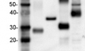
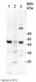
10 µg/well of total protein extracted freshly from Arabidopsis thaliana leaf tissue. All lanes shown are from different Arabidopsis thaliana leaf samples extracted simultaneously. Fresh leaf tissue was ground up directly in 1x Bolt LDS loading buffer (Thermo) and 200mM DTT and denatured at 80°C for 10 min. Samples were separated on 12% SDS-PAGE gel and transferred to nitrocellulose by wet transfer for 1hr at 100V. Blot was blocked with 5 % milk in TBST for 1h/RT with agitation. Blot was incubated with Agrisera Mouse anti-actin monoclonal (AS21 4615) at a dilution of 1:1000, in 5% milk with agitation at 1h RT. The blot was rinsed 3 times for 5 minutes in TBS-T with agitation. Then the membrane was incubated with secondary anti-mouse HRP (Agrisera AS09 627) at 1:10 000 dilution in milk for 1hr at room temp. The blots were washed as above and reaction was visualized using ECL reagent and following manufacture's recommendations. The actin band was visualized after 15 seconds of film exposure.
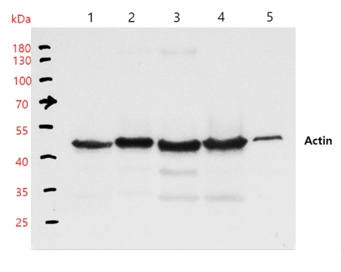
Samples:
1 - 10 µl of Arabidopsis thaliana (Col-0) seedling extract
2 - 10 µl of Nicotiana benthamiana seedlings extract
3 - 10 µl of Nicotiana tabacum leaves extract
4 - 10 µl of Eschscholzia californica leaves extract
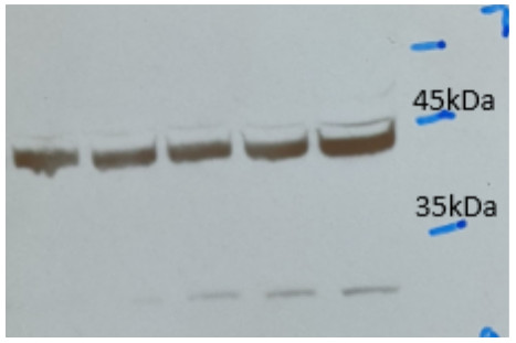
5 - 10 µl of Salvia plebeian hairy roots extractTotal protein was freshly extracted from 50 mg of tissues from species listed above, using 100 µl of 2x SDS sample buffer (0.125M of Tris pH 6.8, 4% SDS, 20% glycerol, 10% 2-mercaptoethanol and 0.2% of bromphenol blue) and denatured with the buffer at 100°C for 3 min. 10 µl of samples were loaded and separated in the 10% SDS-PAGE and blotted for 1h to nitrocellulose (pore size of 0.45 um), using: wet transfer in the cold. Blot was blocked with 3 % milk (PBS-T) for 1h/RT with agitation. Blot was incubated in the primary antibody at a dilution of 1: 10 000/ON with agitation in 3% milk (PBS-T) at 4°C with agitation. The antibody solution was decanted, then washed for 10 min 2 times in PBS-T at RT with agitation. Blot was incubated in matching secondary antibody (Rabbit anti-mouse IgG (H&L), HRP conjugated AS09 627) diluted to 1: 10 000 for 1h/RT with agitation. The blot was washed as above and developed with a chemiluminescent detection reagent. Exposure time was 2 minutes.
Courtesy of Prof. Dr. Sang Un Park's group at Chungnam National University, South Korea
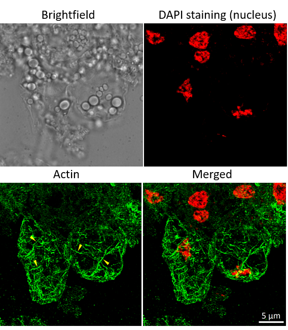
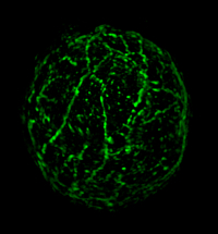
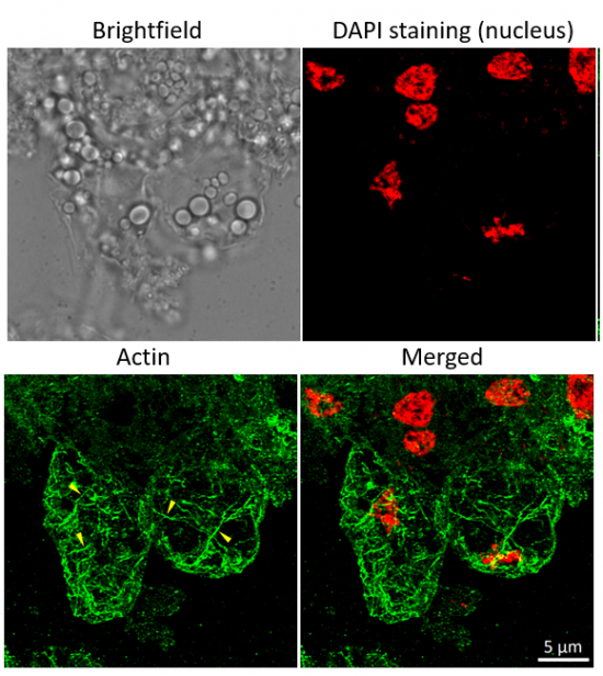
Immunofluorescent localization of actin on suspension culture of Oryza sativa ssp. japonica cv. 'Unggi 9', using anti actin (AS21 4615) and anti-mouse IgG DyLight® conjugated secondary antibodies (AS10 1261). Few representative actin filaments are highlighted by yellow arrowheads. DAPI staining of nuclei is pseudocolored red.
Material: Suspension cultures of Oryza sativa ssp. japonica cv. 'Unggi 9'
Fixation: Packed cell volume to fixer ratio: 250 µl : 5ml
Fixer composition and buffer: 4% (w/v) paraformaldehyde (freshly prepared as 8% stock and 0.2 µm filtered) in Phosphate Buffered Saline (PBS), pH 7.4 (2x stock, 0.2 µm filtered)
Container and method: in 6 cm Petri dish, gentle shaking at room temperature (RT)
Duration: 40 minutes. Triton X100 is not used in fixer. Cells were not shaken during the first 5 mins of fixation to allowed to partially recover from osmotic shock induced by formaldehyde.
Hydrophilization: no
Cell wall digestion: Yes Packed cell volume to enzyme ratio: 100ul : 2ml Enzyme composition: 1.2% Cellulase (chromatically purified, powder, Worthington), 1.2% (R) Pectinase (protease free, liquid, Sigma) Buffer: 0.5% (w/v) MES buffer, pH 5.6
Container and method: in 2 ml microfuge tube by rolling at room temperature (RT)
Duration: 60 minutes
Membrane permeabilization: Triton-X100 (0.35%), 7 min/RT
Antigen retrieval: no
Blocking buffer: Fish gelatin (5% v/v)
Washing buffer: PBS
Primary antibody dilution and incubation time: 1:500, 1hr/RT
Secondary antibody dilution and incubation time and supplier: DyLight® 488 (AS10 1261) 1:600, 45 min/RT
Co-staining of the nucleus (DAPI): Yes
Nucleus staining: 100 ng/ml DAPICourtesy of Dr. Ferhan Ayaydin, Hungarian Centre of Excellence for Molecular Medicine (HCEMM), Szeged, Hungary.
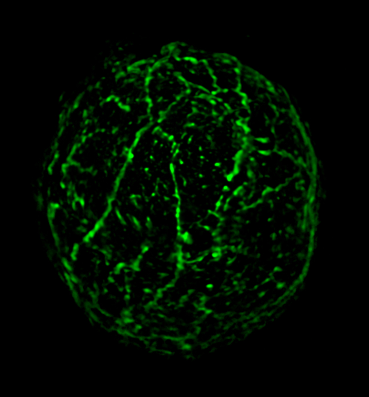
Sample: Maize protoplasts fixed in 4% paraformaldehyde in 1X PBS pH 7.4 for 1 hour and washed 3 times in 1XPBS buffer
Primary antibodies: Agrisera AS21 4615 actin monoclonal; clone 14H4G8;
Secondary antibodies: Donkey anti-Mouse IgG (H&L), DyLight® 488 conjugated AS10 1201 (Agrisera)
Other reagents: 30% Bovine Serum Albumin solution, Sigma Aldrich Cat # A8577-50ML; Phosphate Buffered Saline 10X, Electron Microscopy Sciences Cat # 19342-10; Imaging spacer, Millipore Sigma Grace Bio-Labs SecureSeal™ imaging spacer Cat # GBL654008
Protocol:
- Block isolated protoplasts in a blocking buffer (2% BSA in 1x PBS) for 1 h at room temperature.
- Dilute the primary antibody to 1: 500 in a blocking buffer, mix well, and spin down at 150 x g for 3 min:
- Spin down protoplasts in a blocking buffer at 150 g for 1 -3 min and remove as much supernatant as possible using a pipette tip with a cut end (wide opening).
- Resuspend protoplasts in diluted antibody and incubate for 1 h at room temperature.
- Spin down protoplasts at 150 g for 1 min and remove as much supernatant as possible using a pipette tip with a cut end.
- Wash protoplasts in a blocking buffer 3 times for 5 minutes each. Spin and resuspend as described in step 5 between each washing step.
- Dilute the secondary antibody AS10 1201 to 1: 400, mix well, and spin down at 150 x g for 3 min:
- Incubate protoplasts in diluted antibody for 1 h at room temperature.
- Repeat the washing steps as described in 5-6.
- Resuspend a protoplast pellet in a small volume of PBS 1X, pH 7.4.
- Prepare microscopy slide: attach imaging spacer on top of the slide, peal off top adhesive membrane, add protoplast suspension on to the slide inside the spacer opening, cover with 22x22 mm cover glass No 1.5.
- Imaging: Zeiss Elyra 7 SIM
Courtesy of Courtesy of Dr. Anastasiya Klebanovych, Dr. Kirk Czymmek, Dr. Kevin Cox, Dr. Blake Meyers, at the Donald Danforth Plant Science Center, USA
- Additional Information
-
Additional information: This antibody was produced using CHO cells expression system. Additional information (application): Matching actin protein standard, product number: AS16 4111.
This antibody is compatible with a secondary antibody for fluorescence detection: goat anti-mouse secondary antibody (IRDye 800CW, Li-cor) - Background
-
Background: Actin is a highly conserved protein and an essential component of cell cytoskeleton and plays an important role in cytoplasmic streaming, cell shape determination, cell division, organelle movement and extension growth. Preferentially expressed in young and expanding tissues, floral organ primordia, developing seeds and emerging inflorescence. - Protocols
-
Agrisera Western Blot protocol and video tutorials
Protocols to work with plant and algal protein extracts
Agrisera Educational Posters Collection
- Reviews:
-
zongyu gao | 2022-11-07I used this antibody to detect actin protein from Medicago roots by western blot in 1:5000 dilution. It works quite well, I got specific bands with clean background.
Related products
AS10 1159 | Clonality: Polyclonal | Host: Goat | Reactivity: Mouse IgG (H&L)
AS13 2640 | Clonality: Polyclonal | Host: Rabbit | Reactivity: Actinidia sp., Agostis stoloniferacv. ‘Penncross’,Arabidopsis thaliana, Brassica sp., Cannabis sativa L., Cucumis sativus, Cynara cardunculus, Glycine max, Hordeum vulgare, Nicotiana tabacum, Phaseolus vulgaris, Phoenix dactylifera, Picrorhiza kurroa, Raphanus sativus, Setaria italica, Solanum tuberosum, Triticum aestivum, Zea mays
AS16 4111S | Protein/positive control