1
Anti-GLDP | Glycine decarboxylase P protein
AS20 4370 | Clonality: Polyclonal | Host: Rabbit | Reactivity: Arabidopsis thaliana, Flaveria spp., Neurachne spp.,Oryza sativa, Zea mays
- Product Info
-
Immunogen: KLH-conjugated peptide, highly conserved in Arabidopsis thaliana GLDP1 UniProt:Q94B78, TAIR: At4g33010 and GLDP2 UniProt: O80988, TAIR: At2g26080 Host: Rabbit
Clonality: Polyclonal Purity: Serum Format: Lyophilized Quantity: 50 µl Reconstitution: For reconstitution add 50 µl, of sterile water Storage: Store lyophilized/reconstituted at -20°C; once reconstituted make aliquots to avoid repeated freeze-thaw cycles. Please remember to spin the tubes briefly prior to opening them to avoid any losses that might occur from material adhering to the cap or sides of the tube. Tested applications: Immunolocalization (IL), Western blot (WB) Recommended dilution: 1:50 (IL), 1 : 5000 (WB) Expected | apparent MW: 113 kDa - Reactivity
-
Confirmed reactivity: Arabidopsis thaliana, Flaveria spp., Neurachne spp.,Oryza sativa, Zea mays Predicted reactivity: grasses, non-succulent dicots
Species of your interest not listed? Contact usNot reactive in: Tecticornia spp., Salsola soda - Application Examples
-
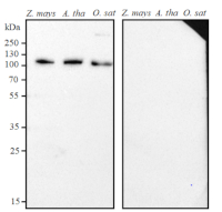
application example
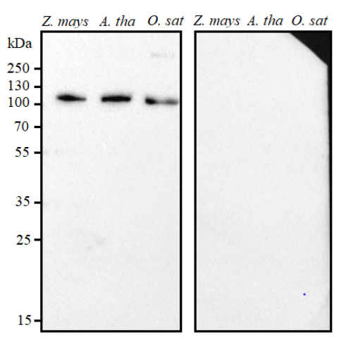
6 µg of total protein extracted freshly from Z. mays = Zea mays, A. tha= Arabidopsis thaliana, O. sat = Oryza sativa, was freshly extracted from leaf tissue in extraction buffer (50 mM HEPES-KOH pH 7.1, 1 mM EDTA pH 8.0, 10 mM DTT, 5 mM MgCl2, 1 mM PMSF, 20 µg/ml chymostatin, 25 µg/ml aprotinin, 1X Protease Inhibitor Cocktail VI, Plant Cell (AG Scientific)). Proteins were denatured with sample buffer (1X = 62.5 mM Tris-HCl pH 6.8, 2% [v/v] SDS, 10% [v/v] glycerol, 0.01% [w/v] bromophenol blue (Sigma), 100 mM DTT), heated to 95°C in a water bath for 3 minutes and collected at 13523 x g in a bench centrifuge. 6µg of protein was loaded per well and separated on a 12% resolving gel (0.375 M Tris-HCl pH 8.8, 12% [v/v] acrylamide, 0.1% [v/v] ammonium persulfate and 0.05% [v/v] TEMED) and 5% stacking gel (0.125 M Tris-HCl pH 6.8, 5% [v/v] acrylamide, 0.1% [v/v] SDS, 0.1% [v/v] ammonium persulfate and 0.025% [v/v] TEMED). Proteins were blotted onto nitrocellulose membrane (pore size 0.45 µm) using Trans-Blot Semi-Dry Transfer Cell (Bio-Rad), and transfer was run at 25 mA per gel stack for 1½ hours. Blots were blocked in blocking solution (5% [w/v] skim milk powder in TBST (12.5 mM Tris, 137 mM NaCl, 2.7 mM KCl, 0.1% [v/v] Tween-20 detergent)) overnight at room temperature with gentle agitation. The blot was rinsed three times quickly in TBST, then incubated in the primary antibody solution at a dilution of 1:5000 in blocking solution for 1 hour at room temperature with gentle agitation. Blots were rinsed three times quickly in TBST, then washed three times for 5 minutes in TBST at room temperature with gentle agitation. Blots were then incubated in secondary antibody solution (goat anti-rabbit IgG horse radish peroxidase (Sigma) diluted 1:3000 in blocking solution) for 1 hour at room temperature with gentle agitation. The blot was washed three times for 5 minutes in TBST at room temperature with gentle agitation, then rinsed twice in TBS (12.5 mM Tris, 137 mM NaCl, 2.7 mM KCl). Blots were developed with chemiluminescent detection reagent, following manufacture's recommendations, incubating at room temperature for 1 minute and imaged with Bio-Rad ChemiDoc system. Exposure time was 40 seconds.Courtesy of Prof. Martha Ludwig, School of Molecular Sciences, Australia
- Additional Information
-
Additional information (application): So far only leaf tissue has been used for immunolocalization and western blot experiments - Background
-
Background: GLDP (Glycine decarboxylase P protein) catalyzes the degradation of glycine. The P protein binds the alpha-amino group of glycine through its pyridoxal phosphate cofactor. Is involved in response to cadmium ion.
Alternative names: AtGLDP1, AtGLDP2, Glycine dehydrogenase (aminomethyl-transferring) 1, Glycine dehydrogenase (aminomethyl-transferring) 2, Glycine cleavage system P protein 1, Glycine cleavage system P protein 2, Glycine decarboxylase 1, Glycine decarboxylase 2, Glycine decarboxylase P-protein 1, Glycine decarboxylase P-protein 2. - Product Citations
-
Selected references: Rakhmankulova et al. (2026). Effect of drought and elevated temperature on the physiological and biochemical properties of C3 and C4 halophytes in Amaranthaceae. Journal of Arid Land Volume 18, Issue 1, January 2026, Pages 131-149.
Khoshravesh et al. (2020). The Evolutionary Origin of C4 Photosynthesis in the Grass Subtribe Neurachninae.Plant Physiol. 2020 Jan;182(1):566-583. doi: 10.1104/pp.19.00925. - Protocols
-
Agrisera Western Blot protocol and video tutorials
Protocols to work with plant and algal protein extracts
Agrisera Educational Poster Collection - Reviews:
-
This product doesn't have any reviews.
Related products

AS09 602 | Clonality: Polyclonal | Host: Goat | Reactivity: Rabbit IgG (H&L)
AS05 074 | Clonality: Polyclonal | Host: Rabbit | Reactivity: A. thaliana,P.hybrida cv. Mitchell, P. grandiflora, S. oleracea, T. aestivum, V. faba | cellular [compartment marker] of mitochondrial matrix
AS06 181 | Clonality: Polyclonal | Host: Rabbit | Reactivity: Arabidopsis thaliana, Catharanthus roseus,Glycine max, Hordeum vulgare, Medicago sativa, Nicotiana tabacum, Oryza sativa, Pisum sativum, Salicornia sp., Silene vulgaris, Scenedesmus quadricauda (algae), Solanum tuberosum, Spinacia oleracea, Triticum turgidum ssp. durum, Zea mays