1
Anti-Cyt f | Cytochrome f protein (PetA) of thylakoid Cyt b6/f-complex (higher plants)
AS20 4377 | Clonality: Polyclonal | Host: Rabbit | Reactivity: Arabidopsis thaliana, Cucumis sativus, Echinola crus-galli, Ficus elastica, Lupinus angustifolius, Nicotiana tabacum, Phaseolus coccineus , Picea abies, Pinus sylvestris, Pisum sativum, Solanum tuberosum, Synechococcus elongatus PCC7942, Zea mays
- Product Info
-
Immunogen: KLH-conjugated peptide derived from Arabidopsis thaliana PetA sequence: UniProt: P56771, TAIR: AtCg00540 Host: Rabbit Clonality: Polyclonal Purity: Immunogen affinity purified serum in PBS pH 7.4. Format: Lyophilized Quantity: 50 µg Reconstitution: For reconstitution add 50 µl, of sterile water Storage: Store lyophilized/reconstituted at -20°C; once reconstituted make aliquots to avoid repeated freeze-thaw cycles. Please remember to spin the tubes briefly prior to opening them to avoid any losses that might occur from material adhering to the cap or sides of the tube. Tested applications: Western blot (WB) Recommended dilution: 1 : 1000 (WB) Expected | apparent MW: 31-32 kDa - Reactivity
-
Confirmed reactivity: Arabidopsis thaliana, Cucumis sativus, Echinola crus-galli, Ficus elastica, Lupinus angustifolius, Nicotiana tabacum, Phaseolus coccineus , Picea abies, Pinus sylvestris, Pisum sativum, Solanum tuberosum, Synechococcus elongatus PCC7942, Zea mays Predicted reactivity: Nicotiana benthamiana
Species of your interest not listed? Contact usNot reactive in: cyanobacteria - Application Examples
-
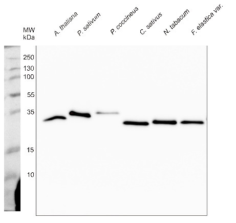
0.25 µg - 1 µg of isolated thylakoids (stored at -80°C): 1 µg of Arabidopsis thaliana thylakoid membranes, 1 µg of Pisum sativum thylakoid membranes, 0.25 µg of Phaseolus coccineus thylakoid membranes, 1 µg of Cucumis sativus thylakoid membranes, 1 µg of Nicotiana tabacum thylakoid membranes, 1 µg of Ficus elastica variegata thylakoid membranes were denatured with 150 µL double diluted Roti®-Load 1 (ROTH, Art.-Nr. K929.1) at 95 °C for 2 min. Samples containing 1 (or 0.25) µg of chlorophyll/well were separated on 14% SDS-PAGE and blotted 45 min at 100V to PVDF (pore size of 0.2 µm) using wet transfer. Blot was blocked with 5% milk 4°C/ON with agitation. Blot was incubated in the primary antibody at a dilution of 1: 1 000 for 3h/RT with agitation in TBS (+0.5% Amersham™ ECL Prime Blocking Agent; cat no: RPN418). The antibody solution was decanted and the blot was washed 2 times for 5 min in TBS-T at RT with agitation. Blot was incubated in Agrisera matching secondary antibody (anti-rabbit IgG horse radish peroxidase conjugated AS09 602) diluted to 1:20 000 in TBS-T (+1% milk) for 1h/RT with agitation. The blot was washed as above and developed for 5 min with Agrisera ECLBright. The Image was recorded using ChemiDoc MP Imaging System (Bio-Rad) with automatic selection of the exposure time.
TBS (25 mM Tris, 500 mM NaCl; pH 7.5) TBS-T (TBS + 0.1% Tween20)
Courtesy of Dr. Radosław Mazur, University of Warsaw, Poland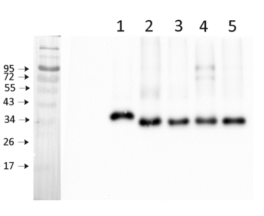
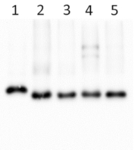
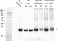
1.0 µg of chlorophyll from chloroplasts of: Pisum sativum (1), Echinochloa crus-galli, M chloroplasts (2), Echinochloa crus-galli, BS (bundle sheath) chloroplasts (3), Zea mays M chloroplasts (4), Zea mays BS (bundle sheath) chloroplasts (5), extracted with 0.4 M sorbitol, 50 mM Hepes NaOH, pH 7.8, 10 mM NaCl, 5 mM MgCl2 and 2 mM EDTA. Samples were denatured with Laemmli buffer at 75ºC for 5 min and were separated on 12% SDS-PAGE and blotted 30 min to PVDF using wet transfer. Blot was blocked with 5% milk for 2h at room temperature (RT) with agitation. Blot was incubated in the primary antibody at a dilution of 1: 1000 overnight at 40ºC with agitation in 1% milk in TBS-T. The antibody solution was decanted and the blot was washed 4 times for 5 min in TBS-T at RT with agitation. Blot was incubated in secondary antibody (anti-rabbit IgG horse radish peroxidase conjugated, from Agrisera, AS09 602, Lot 2001) diluted to 1:20 000 in 1 % milk in TBS-T for 1h at RT with agitation. The blot was washed 5 times for 5 min in TBS-T and 2 times for 5 min in TBS, and developed for 1 min with 1.25 mM luminol, 0.198 mM coumaric acid and 0.009% H2O2 in 0.1 M Tris- HCl, pH 8.5. Exposure time in ChemiDoc System was 73 seconds.
Courtesy of Dr. Wioleta Wasilewska-Dębowska, University of Warsaw, Poland
Samples:
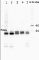
1 - 25 ug of Picea abies total proteins extract obtained from needles using 10% TCA/acetone, pellet was dissolved in 8M Urea, 40 mM Tris-HCl, pH 6.8, 0,1 mM EDTA, 1% SDS
2 - 25 ug of Pinus sylvestris total proteins extract obtained from needles using 10% TCA/acetone, pellet was dissolved in 8M Urea, 40 mM Tris-HCl, pH 6.8, 0,1 mM EDTA, 1% SDS
3 - 10 ug of Lupinus angustifolius thylakoid membranes
4 - 10 ug of Solanum tuberosum thylakoid membranes
5 - 10 ug of Synechococcus elongatus PCC7942 thylakoid membranes
6 - 10 ug of Pisum sativum, thylakoid membranes
MW markers: PageRuler™ Plus Prestained Protein Ladder from ThermoFisher Scientific, cat # 26619.
25 µg/well of total protein from Picea abies (1) and Pinus sylvestris (2) needles extracted with 10% TCA/acetone, and 10 µg/well of chlorophyll from Lupinus angustifolius (3), Solanum tuberosum (4), Synechococcus elongatus PCC7942 (5), and Pisum sativum extracted with 0.4 M sorbitol, 50 mM Hepes NaOH, pH 7.8, 10 mM NaCl, 5 mM MgCl2 and 2 mM EDTA. Samples were denatured with Laemmli buffer at 70°C for 5 min and were separated on 8-16% SDS-PAGE and blotted 40 min to nitrocellulose (pore size of 0.2 um), using semi-dry transfer. Blot was blocked with 5 % milk for 1h at room temperature (RT) with agitation. Blot was incubated in the primary antibody at a dilution of 1: 1 000 for 1h at RT with agitation in 1% milk in TBS-T. The antibody solution was decanted and the blot was rinsed briefly twice, then washed once for 15 min and 3 times for 5 min in TBS-T at RT with agitation. Blot was incubated in secondary antibody (anti-rabbit IgG horse radish peroxidase conjugated, from Agrisera, AS09 602) diluted to 1:25 000 in for 1h/RT with agitation. The blot was washed as above and developed for 5 min with ECL detection kit with iBright FL1500 Imaging System (Thermo Fisher). Exposure time was 745 seconds.
Please note that detection of PetA using this antibody requires optimization of Western blot protocol in terms of protein load and incubation time, depending upon which species is analyzed.
Courtesy of Dr. Elena Pojidaeva Laboratory of Plant Gene Expression Timiryazev Institute of Plant Physiology RAS 127276 Moscow, Russia - Background
-
Background: Multi-subunit complex of cytb6/f is a crucial component for the photosynthetic electron transport chain of higher plants, green algae and cyanobacteria. This complex is catalyzing oxidation of quinols and the reduction the reduction of plastocyanin. This reaction allows to establish the proton force required for the ATP synthesis. Four major subunits build the complex: the petA gene product corresponding to a c-type cytochrome (cytf), the petB gene product corresponding to a b-type/c’-type cytochrome with three haems (cyt b6), the petD gene product (subunit IV, or suIV), and the petC gene product, corresponding to the Rieske/Iron/sulfur protein. - Product Citations
-
Selected references: Penzler et al. (2024). A pgr5 suppressor screen uncovers two distinct suppression mechanisms and links cytochrome b6f complex stability to PGR5. Plant Cell. 2024 Mar 27:koae098. doi: 10.1093/plcell/koae098.
Uflewski et al. (2024). The thylakoid proton antiporter KEA3 regulates photosynthesis in response to the chloroplast energy status. Nat Commun. 2024 Mar 30;15(1):2792. doi: 10.1038/s41467-024-47151-5.
Mu et al. (2024). Plastid HSP90C C-terminal extension region plays a regulatory role in chaperone activity and client binding.Plant J. 2024 Jul 5.doi: 10.1111/tpj.16917.
Lempiainen et al. (2022). Plants acclimate to Photosystem I photoinhibition by readjusting the photosynthetic machinery. Plant Cell Environ. 2022 Oct;45(10):2954-2971. doi: 10.1111/pce.14400. Epub 2022 Aug 16. PMID: 35916195. - Protocols
-
Agrisera Western Blot protocol and video tutorials
Protocols to work with plant and algal protein extracts
Agrisera Educational Poster Collection - Reviews:
-
This product doesn't have any reviews.
Related products

AS09 602 | Clonality: Polyclonal | Host: Goat | Reactivity: Rabbit IgG (H&L)
AS18 4169 | Clonality: Polyclonal | Host: Rabbit | Reactivity: A. thaliana, C. reinhardtii, Echinochloa crus-galli, M. aeruginosa, M. polymorhpa, M. sativa, N. oceanica, Panicum miliaceum, P. sativum, Z. mays
AS05 084 | Clonality: Polyclonal | Host: Rabbit | Reactivity: [global antibody] for higher plants, algae, liverwort, cyanobacteria, diatoms | cellular [compartment marker] of thylakoid membrane
Benefits of using this antibody