4
Anti-H3 | Histone H3 (antigen affinity purified)
AS10 710A | Clonality: Polyclonal | Host: Rabbit | Reactivity: A. thaliana, C. reinhardtii, S. lycopersicum, V. faba, P. patens, S. europaea, Z. mays, Ch. reinhardtii | cellular [compartment marker] of nucleoplasm
- Product Info
-
Immunogen: KLH-conjugated synthetic peptide derived from known H3 sequences, inluding Arabidopsis thaliana H3.3 P59169 (At4g40030,At4g40040,At5g10980), H3.2 P59226(At1g09200,At3g27360,At5g10390,At5g10400,At5g65360), H3-like 2 Q9FXI7 (At1g19890)
Host: Rabbit Clonality: Polyclonal Purity: Immunogen affinity purified serum in PBS pH 7.4. Format: Lyophilized Quantity: 50 µg Reconstitution: For reconstitution add 50 µl of sterile water Storage: Store lyophilized/reconstituted at -20°C; once reconstituted make aliquots to avoid repeated freeze-thaw cycles. Please remember to spin the tubes briefly prior to opening them to avoid any losses that might occur from material adhering to the cap or sides of the tube. Tested applications: ChIp-qPCR (ChIp-qPCR), Immunofluorescence (IF), Western blot (WB) Recommended dilution: 2,5 µg/100 µg of chromatin (ChIp-qPCR), 1: 400 (IF), 1 : 5000 (WB) Expected | apparent MW: 15 | 17 kDa - Reactivity
-
Confirmed reactivity: Arabidopsis thaliana, Cardamine hirsuta, Oryza sativa Predicted reactivity: Brassica oleracea, Capsicum annuum, Chlamydomonas acidophila, Chlamydomonas reinhardtii, Physcomitrium patens, Salicornia europaea, Solanum lycopersicum, Solanum sogarandinum, Solanum tuberosum, Vicia faba, Zea mays Brachypodium distachyon, Brassica napus, Hordeum vulgare, Nicotiana tabacum, Malus domestica, Medicago sativa, Nannochloropsis gaditana, Triticum aestivum, Pinus pinaster, Pisum sativum, Zea mays, Vitis vinifera, Volvox sp.
Species of your interest not listed? Contact usNot reactive in: No confirmed exceptions from predicted reactivity are currently known - Application Examples
-
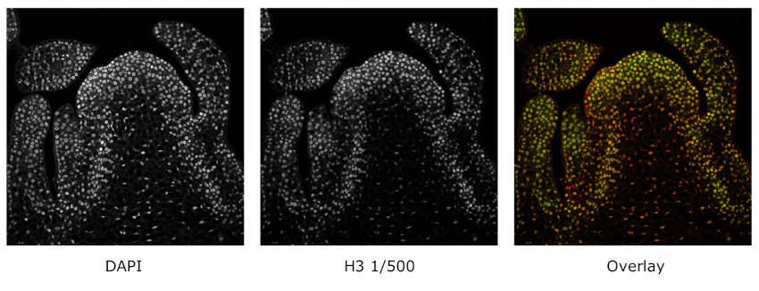
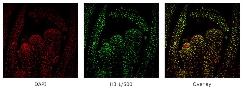
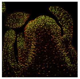
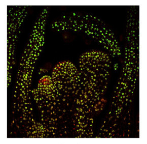
Material: shoots of Arabidopsis thaliana (first set of images) and Cardamine hirsuta (second set of images)
The following protocol was applied:
Fixation: FAA: 4% formaldehyde, 50% ethanol, 5 % acetic acid (A. thaliana)
PFA: 4% formaldehyde in 50 mM phosphate buffer pH 7.5 (C. hirsuta)
Embedding and sectioning: Steedman wax/10 μm sections
Hydrophilization: no
Cell wall digestion: no
Membrane permeabilization: DMSO-IGEPAL
Antigen retrieval: no
Blocking buffer: TSA blocking reagent (Perkin Elmer)
Washing buffer: PBS with 0.1% gelatine
Primary antibody: 1: 500 incubation o/n at 4°C
Secondary antibody: GAR-alexa488 (ThermoFisher) 1:250, 2h/RT
Co-staining of the nucleus (DAPI): yes
Cell wall staining: no
Imaging: Leica Stellaris 5 confocal microscope
Note: fixation performed with: PFA or FAA was giving comparable results.
Dr. Ton Timmers, Max Planck Institute for Plant Breeding Reseach, Cologne, Germany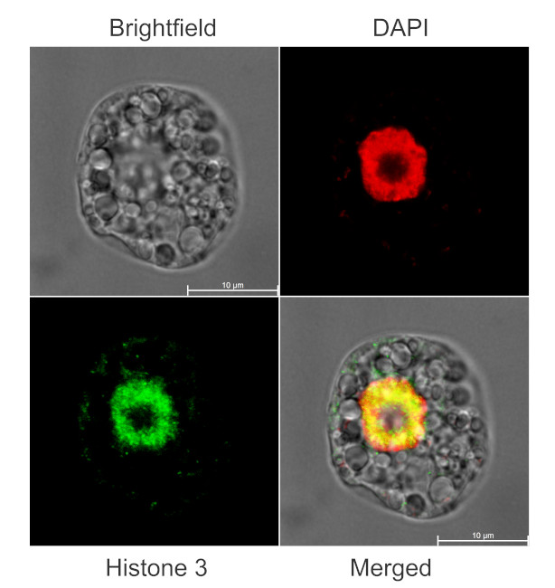
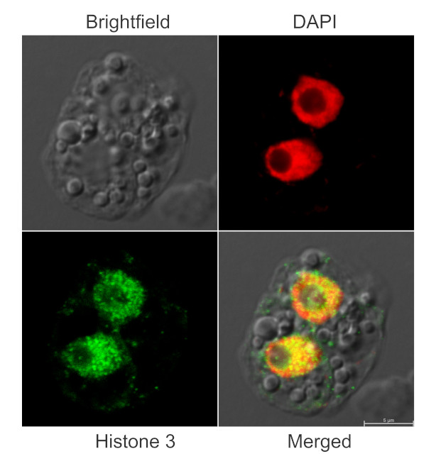
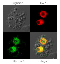
Immunofluorescent localization of Histone 3 on suspension culture of Arabidopsis thaliana (upper image) or Oryza sativa (bottom image), using anti-histone 3 antibodies (AS10 710A) and anti-rabbit IgG DyLight®488 conjugated secondary antibodies (AS10 1165). DAPI staining of nuclei is pseudocolored red.
Material: Suspension cultures of Arabidopsis thaliana, ecotype Landsberg erecta cv.MM1 or Oryza sativa ssp.japonica cv. 'Unggi 9'
Fixation: Packed cell volume to fixer ratio: 250 µl : 5ml
Fixer composition and buffer: 4% (w/v) paraformaldehyde (freshly prepared as 8% stock and 0.2 µm filtered) in Phosphate Buffered Saline (PBS), pH 7.4 (2x stock, 0.2 µm filtered)
Container and method: in 6 cm Petri dish, gentle shaking at room temperature (RT)
Duration: 30 minutes (Arabidopsis thaliana) or 60 minutes (Oryza sativa). Cells were not shaken during the first 5 mins of fixation to allowed to partially recover from osmotic shock induced by formaldehyde.
Hydrophilization: no
Cell wall digestion: Yes
Packed cell volume to enzyme ratio: 100 µl : 2ml Enzyme composition: 1% (A) 1.2% (R) Cellulase (chromatically purified, powder, Worthington) 1% Pectinase (protease free, liquid, Sigma) Buffer: 0.5% (w/v) MES buffer, pH 5.6
Container and method: in 2 ml microfuge tube by rolling at room temperature (RT)
Duration: 30 minutes (Arabidopsis thaliana) or 90 minutes (Oryza sativa)
Membrane permeabilization: Triton-X100 (0.5%), 10 min/RT
Antigen retrieval: no
Blocking buffer: Fish gelatin (5% v/v)
Washing buffer: PBS
Primary antibody dilution and incubation time: 1:400, ON/4ºC
Secondary antibody dilution and incubation time and supplier: anti-rabbit IgG DyLight®488 conjugated secondary antibodies (AS10 1165), 1:600, 1hn/RT
Co-staining of the nucleus (DAPI): Yes
Nucleus staining: 100 ng/ml DAPICourtesy of Dr. Ferhan Ayaydin, Hungarian Centre of Excellence for Molecular Medicine (HCEMM), Szeged, Hungary.
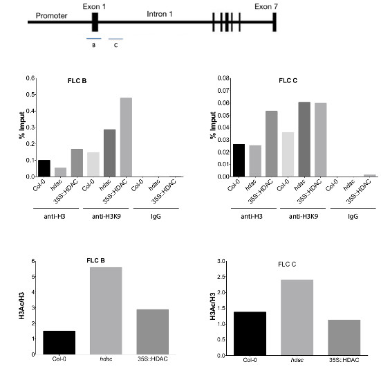
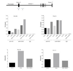
Chromatin Immunoprecipitation: using anti-plant Histone 3 polyclonal antibodies. Chromatin from Arabidopsis thaliana wilde type, deacetylase mutant and over-expressors was cross-linked using formaldehyde. Chromatin was isolated and DNA was sheared along with the bound protein by sonictaion. DNA-protein complex was immunoprecipitated using affinity purified, polyclonal anti-Histone 3 antibodies. Immunoprecipitated DNA was quantified using quantitative PCR and normalized to the input chromatin.
Procedure was according to a protocol described here: Saleh et al. (2008).
Courtesy of Dr. Cristián Holzmann, Catholic University of Chile, Chile - Additional Information
-
Additional information: Cellular [compartment marker] of nucleoplasm, loading control antibody for Chlamydomonas reinhardtii Additional information (application): Protocol for isolation of cytosolic and nuclear fractions can be found here.
Specific protol for ChIP can be found here: Saleh et al. (2008).
- Background
-
Background: Histone 3 (H3) located in nuclei, incorporated into chromatin. Present in nucleosome together with H2A, H2B and H4.
- Product Citations
-
Selected references: Sandhalkar et al. (2025). Photosynthetic acclimation response of Chlamydomonas reinhardtii in synthetic dairy wastewater. J Appl Phycol (2025). https://doi.org/10.1007/s10811-025-03525-w. - Protocols
-
Agrisera Western Blot protocol and video tutorialsAgrisera Western Blot protocol and video tutorials
Protocols to work with plant and algal protein extracts
Agrisera Educational Posters CollectionPreparation of cytosolic and nuclear protein fractions
1. Prepare protoplasts from 50 ml Arabidopsis thaliana cell culture according to the protocol of PEG transfection.
2. Resuspend protoplasts in 10 ml GH buffer and keep the solution on ice for 10 min.
GH buffer: 100mM glycine
0.1% Hexylene glycol
0.37M (4.7% w/v) saccharose
0.3mM Spermine
1.0mM Spermidine
pH 8.3 with Ca(OH)2
3. To release nuclei add Triton X100 to a final concentration of 0.1%. Pipetting gently up and down several
times with a plastic pipette might be necessary to lyse cells.
4. After 5min sediment nuclei by centrifugation at 1000 xg for 15 min at 4°C. Save supernatant as the
cytoplasmic fraction. Wash the pelleted nuclei two times with GHT (GH+0.1% TX100) then finally
resuspended in a suitable volume of extraction buffer + protease inhibitors.Courtesy Dr. Laszlo Bako, Umeå Plant Science Centre
- Reviews:
-
This product doesn't have any reviews.
Related products

AS09 602 | Clonality: Polyclonal | Host: Goat | Reactivity: Rabbit IgG (H&L)

AS09 607 | Clonality: Polyclonal Host: Goat Reactivity: Rabbit IgG (H&L)
AS21 4694 | Clonality: Polyclonal | Host: Rabbit | Reactivity: Negative control IgG for chromatin immunoprecipiation assays (ChIP)