1
Anti-PsbA | D1 protein of PSII, phosphorylated
AS13 2669 | Clonality: Polyclonal | Host: Rabbit | Reactivity: [global antibody] for higher plants
Benefits of using this antibody
- Data sheet
- Product Info
-
Immunogen: KLH-conjugated synthetic peptide derived from available plant PsbA sequences with phosphorylated (T), including Arabidopsis thaliana UniProt: P83755, TAIR:AtCg00020, Oryza sativa P0C434 and other higher plant PsbA sequences
Host: Rabbit Clonality: Polyclonal Purity: Immunogen affinity purified serum in PBS pH 7.4. Format: Lyophilized Quantity: 50 µg Reconstitution: For reconstitution add 50 µl of sterile water Storage: Store lyophilized/reconstituted at -20°C; once reconstituted make aliquots to avoid repeated freeze-thaw cycles. Please remember to spin the tubes briefly prior to opening them to avoid any losses that might occur from material adhering to the cap or sides of the tube. Tested applications: Western blot (WB) Recommended dilution: 1 : 10 000 (WB) Expected | apparent MW: 38 | 28-30 kDa
- Reactivity
-
Confirmed reactivity: Arabidopsis thaliana, Chlamydomonas reinhardtii, Hordeum vulgare, Zea mays Predicted reactivity: Dictos, Conifers, Monocts
Species of your interest not listed? Contact usNot reactive in: Cyanobacteria - Application Examples
-
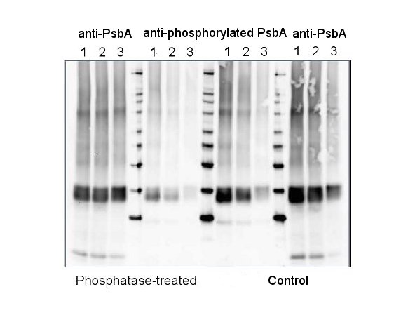
5 µg of total protein from 10 days old seedlings of Hordeum vulgare exposed to high light of 450 µmol to induce phosphorylation (1), or low light of 2 µmol (2), darkness (3) for three hours were extracted with Protein Extraction Buffer PEB (AS08 300). All three samples were treated with lambda protein phosphatase (NEB) to dephosphorylate the PsbA protein (Phosphatase-treated). Samples were diluted with 1X sample buffer (NuPAGE LDS sample buffer (Invitrogen) supplemented with 50 mM DTT and heated at 70°C for 5 min and keep on ice before loading. Protein samples were separated on NuPAGE 4-12% Tris-Bis gel (Invitrogen) LDS-PAGE and blotted for 1h to 1.5h on PVDF using tank transfer. Blots were blocked immediately following transfer in 2% blocking reagent or 5% non-fat milk dissolved in 20 mM Tris, 137 mM sodium chloride pH 7.6 with 0.1% (v/v) Tween-20 (TBS-T) for 1h at room temperature with agitation. Blots were incubated in the primary antibody anti-PsbA (AS05 084, first 3 and last 3 lanes on a blot) and anti-phosphorylated PsbA (AS13 2669, middle lanes) at a dilution of 1: 10 000 (in blocking reagent) for 1h at room temperature with agitation. The antibody solution was decanted and the blot was rinsed briefly twice, and then washed 1x15 min and 3x5 min with TBS-T at room temperature with agitation. Blots were incubated in secondary antibody (Agrisera anti-rabbit IgG horse radish peroxidase conjugated, recommended secondary antibody AS09 602) diluted to 1:25 000 in blocking reagent for 1h at room temperature with agitation. The blots were washed as above. The blot was developed for 5 min with extreme femtogram range chemiluminescnce detection reagent according the manufacturers instructions. Images of the blots were obtained using a CCD imager (FluorSMax, Bio-Rad) and Quantity One software (Bio-Rad). Exposure time was 2 minutes. - Additional Information
-
Additional information (application): This antibody is detecting phosphorylated PsbA protein.
Antibodies are purified on a non-phosphorylated peptide. - Background
-
Background: The psbA gene has been cloned from many species of plants, green algae, and cyanobacteria. The psbA gene is located in the chloroplast genome and encodes for the D1 protein, a core component of Photosystem II. PsbA/D1 is rapidly cycled under illumination in all oxygenic photobionts. Tracking PsbA pools using the Global PsbA antibody can show the functional content of Photosystem II in a wide range of samples.
Alternative names: 32 kDa thylakoid membrane protein, photosystem II protein D1. - Product Citations
-
Selected references: Pilarska et al. (2025). Salinity-induced changes in the PSII/LHCII phosphorylation and organization of the photosynthetic protein complexes in the halophyte Mesembryanthemum crystallinum L. Journal of Plant Physiology, 10 July 2025, 154567.
Wójtowicz et al. (2025). Shrink or expand? Just relax! Bidirectional grana structural dynamics as early light-induced regulator of photosynthesis. New Phytol . 2025 Jun;246(6):2580-2596. doi: 10.1111/nph.70175.
Collombat et al. (2025). Arabidopsis conditional photosynthesis mutants abc1k1 and var2 accumulate partially processed thylakoid preproteins and are defective in chloroplast biogenesis. Commun Biol . 2025 Jan 22;8(1):111. doi: 10.1038/s42003-025-07497-y.
Verhoeven and Kornkven (2023). Differences in photoprotective strategy during winter in Eastern white pine and white spruce. Tree Physiol. 2023 Oct 20:tpad131.doi: 10.1093/treephys/tpad131.
Mazur et al. (2021) The SnRK2.10 kinase mitigates the adverse effects of salinity by protecting photosynthetic machinery. Plant Physiol. 2021 Dec 4;187(4):2785-2802. doi: 10.1093/plphys/kiab438. PMID: 34632500; PMCID: PMC8644180.
Upadhyaya and Jagadeeshwar Rao (2019). Reciprocal regulation of photosynthesis and mitochondrial respiration by TOR kinase in Chlamydomonas reinhardtii. Plant Direct Volume 3, Issue 11.
Xing et al. (2017). Deletion of CGLD1 Impairs PSII and Increases Singlet Oxygen Tolerance of Green Alga Chlamydomonas reinhardtii. Front. Plant Sci., 15 December 2017. https://doi.org/10.3389/fpls.2017.02154.
Li et al. (2015). Effect of hydrogen sulfide on D1 protein in wheat under drought stress. Acta Physiologiae Plantarum November 2015, 37:225. - Protocols
-
Agrisera Western Blot protocol and video tutorials
- Reviews:
-
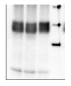
Radoslaw Mazur | 2019-04-29Plant species: Arabidopsis thaliana
Technique: western-blot. The antibody recognizes D1-P as a double band.
Load per well: 1 ug of chlorophyll
Dilution: 1:10000
Secondary antibody: Agrisera AS09 602, dilution 1:10000 (antibody stock in 50% glycerol).
Detection: chemiluminescence
Related products

AS09 602 | Clonality: Polyclonal | Host: Goat | Reactivity: Rabbit IgG (H&L)

AS09 607 | Clonality: Polyclonal Host: Goat Reactivity: Rabbit IgG (H&L)
AS05 084 | Clonality: Polyclonal | Host: Rabbit | Reactivity: [global antibody] for higher plants, algae, liverwort, cyanobacteria, diatoms | cellular [compartment marker] of thylakoid membrane
Benefits of using this antibody