1
Anti-SAM1-4 | S-adenosylmethionine synthase
AS16 3148 | Clonality: Polyclonal | Host: Rabbit | Reactivity: Arabidopsis thaliana
- Product Info
-
Immunogen: Host: Rabbit Clonality: Polyclonal Purity: Serum Format: Lyophilized Quantity: 50 µl Reconstitution: For reconstitution add 50 µl of sterile water Storage: Store lyophilized/reconstituted at -20°C; once reconstituted make aliquots to avoid repeated freeze-thaw cycles. Please remember to spin the tubes briefly prior to opening them to avoid any losses that might occur from material adhering to the cap or sides of the tube. Tested applications: Western blot (WB) Recommended dilution: 1 : 3000 (WB) Expected | apparent MW: 43.2 | 45 kDa (Arabidopsis thaliana)
- Reactivity
-
Confirmed reactivity: Arabidopsis thaliana, Malus domestica, Raphanus sativus Predicted reactivity: Beta vulgaris, Brassica sp., Citrus sp., Coffea canephora, Caomelina sativa, Capsella rubella, Cucumis melo, Cucumis sativus, Genlisea aurea, Gentiana triflora, Guzmania wittmacki, Gossypium raimondii, Eucalyptus grandis, Eutrema salsugineum, Ipomoea batatas, Jatropha curcas, Musa acuminata, Nicotiana sp., Oryza brachyantha, Phoenix dactylifera, Populus sp., Prunus sp., Ricinus communis, Sesamum indicum, Setaria italica, Solanum pennellii, Solanum tuberosum, Spinacia oleracea, Tarenaya hassleriana, Theobroma cacao, Zea mays
Species of your interest not listed? Contact usNot reactive in: Prunus domestica - Application Examples
-
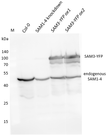
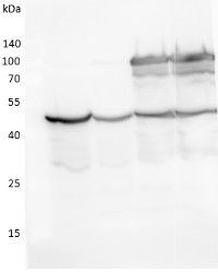
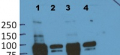
70 μg of total protein from Arabidopsis thaliana wt Col-0, SAM1-4 knockdown, and two oeSAM3-YFP lines extracted with mortar and pestle using 2xSDS loading buffer (100 mM Tris-HCl pH 6.8, 4% SDS, 0.02% bromophenol blue, 200 mM DTT), and denatured in the same buffer at 95°C for 10 min. Samples were separated on 11% SDS-PAGE and blotted for 1h to PVDF using tank transfer. Blots were blocked with 5% milk powder in TBS-T overnight in a cold room with agitation. Blot was incubated in the primary antibody at a dilution of 1: 1 000 for 1h at RT with agitation. The antibody solution was decanted and the blot was rinsed five times for 4 min in TBS-T at RT with agitation. Blot was incubated in secondary antibody (anti-rabbit IgG HRP-conjugated, from Agrisera) diluted to 1: 20 000 in for 2h at RT with agitation. The blot was washed as above and developed for 2 min with chemiluminescent detection reagent. The proteins were detected using CCD Image Fusion Fx7 after 60 seconds exposure time.
Courtesy of Louis-Valentin Meteignier, LGBP, Faculté des Sciences de Luminy, France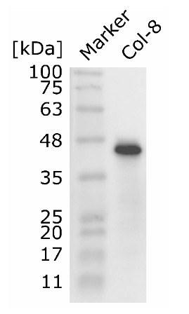
Leaf tissue of Arabidopsis thaliana Col-8 plants was disrupted via mortar and pestle under liquid nitrogen. Leaf protein was extracted by addition of appropriate amounts of extraction buffer (6 M Guanidine-HCl, 100 mM HEPES, 5 mM EDTA and 1x Halt™ Protease Inhibitor-Cocktail). Extracted protein was chloroform/methanol-precipitated and resolved in 2 % SDS, 50 mM Tris (pH 7.4). Protein concentration was determined using BCA assay. 100 µg of cleaned protein were mixed with 4x LDS sample buffer (final conc. 1x), 50 mM DTT (final) and heated 2x to 95 °C for 5 minutes for denaturation. 2.5 µg of protein were loaded per lane on a 12 % SDS PAGE gel. After separation, proteins were blotted to a PVDF membrane for 30 minutes with 25 V and 1 A in a semi-dry blot. Blots were blocked with 5 % Milk in TBS (= Tris buffered saline) for 2 h at room temperature. Blots were subsequently washed with TBS-T (TBS + 0.05 % Tween20) for 1h at room temperature with 3 exchanges of wash buffer. Primary antibody (anti SAM1-4) was used at a dilution of 1:2000 in 1 % Milk in TBS-T and blots were incubated over night at 4 °C with gentle agitation. Primary antibody was decanted and blots were again washed for 1 h with TBS-T as mentioned above. Secondary antibody (anti Rabbit IgG, HRP conjugated) was used at a dilution of 1:20000 in 1 % Milk in TBS-T and blots were incubated for 1 h at room temperature. After decanting of secondary antibody, blots were again washed with TBS-T for 1h. Before Chemiluminescence detection, blots were shortly washed with TBS without Tween20 3x 5 minutes at room temperature. Chemiluminescent detection reagent was used for signal detection. Images of the blots were obtained using ChemiDoc™ XRS (Bio-rad) in high resolution mode. Exposure time was 20 seconds.
Courtesy of phd student Andreas Perrar, Prof. Dr. Pitter Huesgen group, Forschungszentrum Jülich, Germany - Background
-
Background: SAM (S-adenosylmethionin synthetase) is an enzyme which catalyzes the formation of S-adenosylmethionine from methionine and ATP. The overall synthetic reaction is composed of two sequential steps. It is a donor of methyl groups in methylation reactions, and is involved in a number of processes such as: response to cadmium ions and salt stress, as well as, ethylene and lignin biosynthesis. - Product Citations
-
Selected references: Piechowiak et al. (2025), Ozone Treatment Enhances Antioxidant Status and Energy Metabolism in Radish (Raphanus sativus) Sprouts. Springer Nature, Food Bioprocess Technology. - Reviews:
-
Pitter Huesgen | 2020-11-16Showed a strong single 45 kDa band in Arabidopsis leave tissue. Recommended load was to high for us, 2.5 ug total proteome were sufficient.
Detailed protocol:
Leaf tissue of Arabidopsis thaliana Col-8 plants was disrupted via mortar and pestle under liquid nitrogen. Leaf protein was extracted by addition of appropriate amounts of extraction buffer (6 M Guanidine-HCl, 100 mM HEPES, 5 mM EDTA and 1x Halt™ Protease Inhibitor-Cocktail). Extracted protein was chloroform/methanol-precipitated and resolved in 2 % SDS, 50 mM Tris (pH 7.4). Protein concentration was determined using BCA assay. 100 µg of cleaned protein were mixed with 4x LDS sample buffer (final conc. 1x), 50 mM DTT (final) and heated 2x to 95 °C for 5 minutes for denaturation. 2.5 µg of protein were loaded per lane on a 12 % SDS PAGE gel. After separation, proteins were blotted to a PVDF membrane for 30 minutes with 25 V and 1 A in a semi-dry blot. Blots were blocked with 5 % Milk in TBS (= Tris buffered saline) for 2 h at room temperature. Blots were subsequently washed with TBS-T (TBS + 0.05 % Tween20) for 1h at room temperature with 3 exchanges of wash buffer. Primary antibody (anti SAM1-4) was used at a dilution of 1:2000 in 1 % Milk in TBS-T and blots were incubated over night at 4 °C with gentle agitation. Primary antibody was decanted and blots were again washed for 1 h with TBS-T as mentioned above. Secondary antibody (anti Rabbit IgG + HRP) was used at a dilution of 1:20000 in 1 % Milk in TBS-T and blots were incubated for 1 h at room temperature. After decanting of secondary antibody, blots were again washed with TBS-T for 1h. Before Chemiluminescence detection, blots were shortly washed with TBS without Tween20 3x 5 minutes at room temperature. Chemiluminescent detection reagent was used for signal detection. Images of the blots were obtained using ChemiDoc™ XRS (Bio-rad) in high resolution mode. Exposure time was 20 seconds.
Related products

AS09 602 | Clonality: Polyclonal | Host: Goat | Reactivity: Rabbit IgG (H&L)
AS16 3148A | Clonality: Polyclonal | Host: Rabbit | Reactivity: Arabidopsis thaliana
AS12 1865 | Clonality: Polyclonal | Host: Rabbit | Reactivity: Arabidopsis thaliana