1
Anti-SRK2E | Ser/Thr-protein kinase SnRK2,6
AS13 2635 | Clonality: Polyclonal | Host: Rabbit | Reactivity: Arabidopsis thaliana
- Product Info
-
Immunogen: KLH-conjugated inique synthetic peptide derived from Arabidopsis thaliana SRK2E sequence UniProt: Q940H6, TAIR: AT4G33950
Host: Rabbit Clonality: Polyclonal Purity: Immunogen affinity purified serum in PBS pH 7.4. Format: Lyophilized Quantity: 50 µg Reconstitution: For reconstitution add 50 µl of sterile water Storage: Store lyophilized/reconstituted at -20°C; once reconstituted make aliquots to avoid repeated freeze-thaw cycles. Please remember to spin the tubes briefly prior to opening them to avoid any losses that might occur from material adhering to the cap or sides of the tube. Tested applications: Western blot (WB), pull-down assay, Immunoprecipitation (IP) Recommended dilution: 5 µg (IP), 1 : 1000 (WB) 10 µg (pull-down assay) Expected | apparent MW: 41 kDa
- Reactivity
-
Confirmed reactivity: Arabidopsis thaliana Predicted reactivity: Hordeum vulgare
Species of your interest not listed? Contact us
Not reactive in: No confirmed exceptions from predicted reactivity are currently known - Application Examples
-
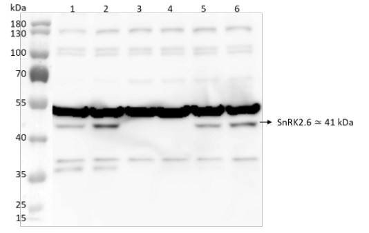
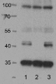
Samples:
1 - 50 µg of Arabidopsis thaliana Col0 mock-treated (MG132 50 µM, 6 hours)
2 - 50 µg of Arabidopsis thaliana Col0 ABA-treated (MG132 50 µM + ABA 50 µM, 6 hours)
3 - 50 µg of Arabidopsis thaliana ost1(snrk2.6) mock-treated (MG132 50 µM, 6 hours)
4 - 50 µg of Arabidopsis thaliana ost1(snrk2.6) ABA-treated (MG132 50 µM + ABA 50µM 6, hours)
5 - 50 µg of Arabidopsis thaliana abi1-2 mock-treated (MG132 50 µM, 6 hours)
6 - 50 µg of Arabidopsis thaliana abi1-2 ABA-treated (MG132 50 µM + ABA 50 µM, 6 hours)
The ost1-3 (SALK_008068) and the abi1-2 (SALK_72009) mutants were used as controls.50 µg/well of total protein extracted freshly from Arabidopsis thaliana roots with extraction buffer containing: 150 mM NaCl, 50 mM Tris-HCL pH 8, 1% Triton X-100, anti-proteases cocktail (Complete mini EDTA free, “ROCHE”) (1 tablet for 10ml), 3 mM DTT, 50 mM MG132, or 50 mM ABA and denatured with exact buffer components at 95 °C/5 min. Samples were separated on 10% SDS-PAGE and blotted overnight (ON) to PVDF (Inmobilon®-FL) (pore size of 0.45 µm), using: wet transfer. Blot was blocked with 3% milk for: 6h/RT with agitation. Blot was incubated in the primary antibody at a dilution of 1: 10 000 in TBS-T 1X for ON/4°C with agitation. The antibody solution was decanted, and the blot was rinsed briefly twice, then washed 3 times for 5 min in TBS-T at RT with agitation. Blot was incubated in matching secondary antibody (Goat anti-rabbit IgG HRP conjugated, AS09 602, Agrisera) diluted to 1: 10000 in for 1h/RT with agitation. The blot was washed as above and developed with a following chemiluminescent detection reagent: AgriseraECL SuperBright (AS16 ECL-S-10, Agrisera). Exposure time was 30 seconds.
Courtesy of Drs. Javier Ocaña, Alberto Coego and Pedro L. Rodriguez, CSIC, Spain
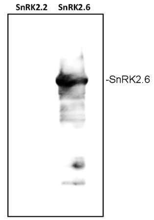
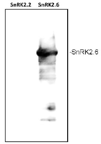
Bacterial lysates were separated on 12% SDS-PAGE and blotted 1h to PVDF using semi-dry or tank transfer. Blots were blocked with 5 % milk for 1h at room temperature (RT) with agitation. Blot was incubated in the primary antibody at a dilution of 1: 3 000 for 1h at RT with agitation. The antibody solution was decanted and the blot was rinsed briefly twice, then washed once for 15 min and 3 times for 5 min in PBS-T at RT with agitation. Blot was incubated in secondary antibody (anti-rabbit IgG horse radish peroxidase conjugated, AS09 602 from Agrisera) diluted to 1:50 000 in for 30 min. at RT with agitation. The blot was washed as above and developed for 3 min with ECL according to the manufacturer's instructions. Exposure time was 30 seconds.
Courtesy of Dr. Agnieszka Ludwików, UAM, Poznań, Poland
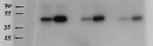
Protein A agarose beads (40µl) where coated with 10µl (1µg/ul) antibodies and after incubation with amount of extract (10 mg/ml) indicated washed extensively and loaded on gel. In gel kinase assay was performed as described in Fujii, 2007.
Autoradiograph shows immunoprecipitated kinase from plant extracts. 1 beads with BSA 20 µl loaded on gel 2 beads with plant extract (WT) 20 µl loaded on gel 3 beeds with plant extract (mutant X) 20 µl loaded on gel 4 beads with BSA 10 µl loaded on gel 5 beads with plant extract (WT) 10 µl loaded on gel 6 beeds with plant extract (mutant X) 10 µl loaded on gel 7 beads with BSA 5 µl loaded on gel 8 beads with plant extract (WT) 5 µl loaded on gel 9 beeds with plant extract (mutant X) 5 µl loaded on gel
Courtesy of Dr. Szymon Świeżewski, Institute of Biochemistry and Biophysics, Polish Academy of Science, Warsaw, Poland - Background
-
Background: SRK2E (Serine/threonine-protein kinase SRK2E) is an activator of the abscisic acid (ABA) signaling pathway, regulating such ABA responses as: stomato closure, response to drought and plant pathogens.
Synonyms: OST1, Open stomata 1 - Product Citations
-
Selected references: Wu et al. (2026). H2S-modulated alternative splicing of the protein phosphatase gene HAB2 drives divergent stomatal dynamics in Arabidopsis. Journal of Experimental Botany, erag036, doi.org/10.1093/jxb/erag036.
Wang et al. (2017). Reciprocal Regulation of the TOR Kinase and ABA Receptor Balances Plant Growth and Stress Response. Mol Cell. 2017 Dec 27. pii: S1097-2765(17)30930-9. doi: 10.1016/j.molcel.2017.12.002. - Protocols
-
Agrisera Western Blot protocol and video tutorials
Protocols to work with plant and algal protein extracts
Agrisera Educational Posters Collection - Reviews:
-
Anna Kulik | 2023-03-22Antibodies work fine for WB.We were able to detect strong bands for SnRK2.6 in Arabidopsis thaliana leaves. We used 25 ug of total protein and the protocol recommended by Agrisera. In roots, an extremely low signal was observed.
Related products

AS09 602 | Clonality: Polyclonal | Host: Goat | Reactivity: Rabbit IgG (H&L)
AS14 2783 | Clonality: Polyclonal | Host: Rabbit | Reactivity: Arabidopsis thaliana