Visar bild 1 av
1
Anti-TGG1 | Myrosinase 1 ( BGL38)
Product no: AS20 4416
AS20 4416 | Clonality: Polyclonal | Host: Rabbit | Reactivity: Arabidopsis thaliana
380
€
Customer reviews
Delivery:
3-6 business days
- Product Info
-
Immunogen: BSA-conjugated peptide, derived from N-terminus of Arabidopsis thaliana TGG1, UniProt: P37702, TAIR: At5g26000 Host: Rabbit Clonality: Polyclonal Purity: Total IgG. Protein A purified. Format: Liquid at 2 mg/ml, in PBS with 50% glycerol. Quantity: 200 µg Storage: Store at -20°C; once reconstituted make aliquots to avoid repeated freeze-thaw cycles. Please remember to spin the tubes briefly prior to opening them to avoid any losses that might occur from material adhering to the cap or sides of the tube. Tested applications: ELISA (ELISA), Immunogold (IG), Immunohistochemistry (IHC), Western blot (WB) Recommended dilution: Assay dependent (ELISA), 1: 1000 - 1: 2500 (IG), 1: 500 - 1: 1000 (IHC), 1: 1000-1: 3000 (WB) Expected | apparent MW: 61 | 77 kDa - Reactivity
-
Confirmed reactivity: Arabidopsis thaliana Predicted reactivity: Species of your interest not listed? Contact us Not reactive in: No confirmed exceptions from predicted reactivity are currently known - Application Examples
-
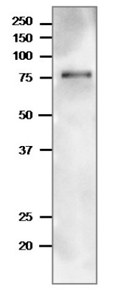
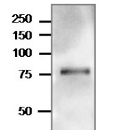
Arabidopsis thaliana total leaf was freshly extracted with 2x SDS-sample buffer (+ 2ME) for SDS-PAGE and denatured with 4X SDS buffer at 95°C for 5 min. 10 µg of protein was loaded and separated on 15-20 % SDS-PAGE and blotted 1h to PVDF membrane. Blot was blocked with 3 % skim milk/TBS-T, 1h/RT with agitation. Blot was incubated in the primary antibody at a dilution of 1: 1000 in TBS-T for 1h/RT. The antibody solution was decanted and the blot was washed 4 times for 10 min in TBS-T at RT with agitation. Blot was incubated in matching secondary antibody (anti-rabbit IgG horse radish peroxidase conjugated) diluted to 1:10 000 in for 1h/RT with agitation. The blot was washed as above and developed with a chemiluminescent detection reagent, following manufacture's recommendations.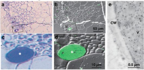
Immunolocalization of TGG1 in sections of Arabidopsis thaliana 48 days-old rosette leaves. Panel to the left: CBB staining (a,c). Middle panel: Reaction with anti-TGG1 antibodies used at 1: 1000 dilution and followed by visualization with AlexaFluor488 goat anti-rabbit antibodies at 1: 1000 (b.d). Right panel: electron microscopy of ultrathin sections mounted on Formvar-coated nickle grid. Anti-TGG1 antibodies were used at 1: 1000 dilution and following washing in PBS the sections were incubated with anti-rabbit IgG conjugated gold particles (AuroProbe EM). CW- cell wall, V- vacuole.
Protocol:
Rosette leaves of Arabidopsis thaliana were fixed with 4% (w/v) paraformaldehyde and 1% glutaraldehyde in 0.05 M cacodylate buffer (pH 7.4) at 4°C for 3 h. After washing with 0.02 M cacodylate buffer (pH 7.4), these tissues were dehydrated with acetone and embedded in LR white resin at –20°C. Sections were cut on an ultramicrotome (Leica, Reichert Division, Vienna, Austria) for both light microscopic and electron microscopic analyses.
- Additional Information
-
Additional information (application): 19 amino acids of transit peptide are not present in the mature protein - Background
-
Background: In Brassicaceae, the enzyme myrosinase (beta-thioglucoside glucohydrolase, TGG) degrades glucosinolates to produce toxins like thiocyanates, isothiocyanates, nitriles, epithionitriles or oxazolidine- 2-thiones that deter herbivory. There are two TGG enzymes, TGG1 and TGG2, which have a redundant function. Cellular localization: vacuole. - Product Citations
-
Selected references: Farid et al. (2011). Arabidopsis thaliana alpha1,2-glucosyltransferase (ALG10) is required for efficient N-glycosylation and leaf growth. Plant J. 2011 Oct;68(2):314-25.doi: 10.1111/j.1365-313X.2011.04688.x. (Western blot)
Shirakawa et al. (2010). Arabidopsis Qa-SNARE SYP2 proteins localized to different subcellular regions function redundantly in vacuolar protein sorting and plant development. Plant J. 2010 Dec;64(6):924-35.doi: 10.1111/j.1365-313X.2010.04394.x. (Western blot)
Ueda et al. (2006). AtVAM3 is required for normal specification of idioblasts, myrosin cells. Plant Cell Physiol. 2006 Jan;47(1):164-75. doi: 10.1093/pcp/pci232. (Immunlocalization, Western blot). - Protocols
-
Agrisera Western Blot protocol and video tutorials
Protocols to work with plant and algal protein extracts
Agrisera Educational Posters Collection - Reviews:
-
This product doesn't have any reviews.
Related products

AS09 602 | Clonality: Polyclonal | Host: Goat | Reactivity: Rabbit IgG (H&L)
219 €
AS12 1858 | Clonality: Polyclonal | Host: Rabbit | Reactivity: Arabidopsis thaliana, Solanum lycopersicum
329 €
AS12 1859 | Clonality: Polyclonal | Host: Rabbit | Reactivity: Arabidopsis thaliana
329 €