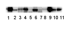
| So-called "Garden blot" means that the same antibody is used on samples from wide taxonomic range. In the presented example, samples were extracted from dicotyl plants, cyanobacteria, diatoms and algae. On the sequence level, the peptide which was used to elicit anti-RbcL antibody, AS03 037 is perfectly conserved in RbcL from all analyzed species. However, clear differences in RbcL band intensity can be observed. This difference can be attributed to:
|
|
Technical tips
Latest
Are blocking conditions nothing to pay attention to?2026-06-18 What are the most common misconceptions about primary antibodies?
2026-05-27 What are the most common mistakes done with secondary antibodies?
2026-05-26 Simple extraction protocol for membrane proteins
2026-04-17 How to improve signal with a weak antibody?
2026-03-26 Same antibody, different outcome on samples from wide taxonomic range
2026-02-23 Can the quality of protein transfer be checked before the blot is developed with antibodies?
2026-01-16 Is wild type a wild type?
2025-12-29 Why an antibody may detect tagged protein but not endogenous one and in some cases endogenous protein but not its tagged version?
2025-10-30 Calclulated and aparent molecular weight of detected protein is different, why?
2025-10-09
Archive
- June - 2026
- May - 2026
- April - 2026
- March - 2026
- February - 2026
- January - 2026
- December - 2025
- October - 2025
- September - 2025
- August - 2025
- July - 2025
- June - 2025
- May - 2025
- April - 2025
- March - 2025
- February - 2025
- January - 2025
- December - 2024
- November - 2024
- September - 2024
- July - 2024
- June - 2024
- May - 2024
- March - 2024
- February - 2024
- December - 2023
- November - 2023
- September - 2023
- July - 2023
- May - 2023
- March - 2023
- January - 2023
- December - 2022
- November - 2022
- October - 2022
- September - 2022
- August - 2022
- June - 2022
- May - 2022
- March - 2022
- February - 2022
- January - 2022
- November - 2021
- October - 2021
- August - 2021
- June - 2021
- May - 2021
- April - 2021
- March - 2021
- February - 2021
- January - 2021
- December - 2020
- November - 2020
- October - 2020
- September - 2020
- August - 2020
- July - 2020
- June - 2020
- May - 2020
- April - 2020
- March - 2020
- January - 2020
- November - 2019
- October - 2019
- March - 2019
- April - 2017
- February - 2017
- May - 2016
- February - 2014
- September - 2013
- December - 2010