1

Donkey anti-Rabbit IgG (H&L), DyLight® 488 conjugated, min, cross-reactivity to bovine, chicken, goat, guinea pig, hamster, horse, human, mouse, rat, sheep IgG
AS10 1165 | Clonality: Polyclonal | Host: Donkey | Reactivity: Rabbit IgG (H&L)
- Product Info
-
Immunogen: Purified Rabbit IgG, whole molecule
Host: Donkey Clonality: Polyclonal Purity: Immunogen affinity purified donkey IgG. Format: Lyophilized Quantity: 1 mg Reconstitution: For reconstitution add 1.1 ml of sterile water. Let it stand 30 minutes at room temperature to dissolve. Prepare fresh working dilutions daily Storage: Store lyophilized material at 2-8°C. Product is stable for 4 weeks at 2-8°C after rehydration. For long time storage after reconstitution, dilute the antibody solution with glycerol to a final concentration of 50% glycerol and store as liquid at -20°C, to prevent loss of enzymatic activity. For example, if you have reconstituted 1 mg of antibody in 1.1 ml of sterile water add 1.1 ml of glycerol. Such solution will not freeze in -20°C, If you are using a 1:5000 dilution prior to diluting with glycerol, then you would need to use a 1:2500 dilution after adding glycerol. Prepare working dilution prior to use and then discard. Be sure to mix well but without foaming. Recommended dilution: 1 : 20-1 : 2000 for most applications - Application Examples
-
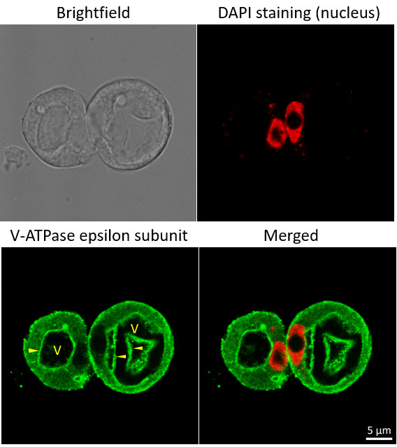
Immunofluorescent localization of V-ATPase epsilon subunit of tonoplast H+ATPase in suspension culture of Oryza sativa ssp. japonica cv. 'Unggi 9', using goat anti-V-ATPase, epsilon subunit of tonoplast antibodies (AS07 213) and donkey anti-rabbit IgG, DyLight® 488 conjugated (AS10 1165, Agrisera). Vacuolar membrane, tonoplast, is highlighted by yellow arrowheads. DAPI staining of nuclei is pseudocolored red.
Method
Material: Suspension cultures of Oryza sativa ssp. japonica cv. 'Unggi 9
Fixation: Packed cell volume to fixer ratio: 250 µl : 5ml
Fixer composition and buffer: 4% (w/v) paraformaldehyde (freshly prepared as 8% stock and 0.2 µm filtered) 0.01% (v/v) Triton-X100 in Phosphate Buffered Saline (PBS), pH 7.4 (2x stock, 0.2 µm filtered) Container and method: in 6 cm Petri dish, gentle shaking at room temperature (RT) Duration: 40 min
Hydrophilization: No
Cell wall digestion: Yes Packed cell volume to enzyme ratio: 100ul : 2ml Enzyme composition: 1% (A) 1.2% (R) Cellulase (chromatically purified, powder, Worthington) 1% (A) 1.2% (R) Pectinase (protease free, liquid, Sigma) Buffer: 0.5% (w/v) MES buffer, pH 5.6 Container and method: in 2 ml microfuge tube by rolling at room temperature (RT)
Duration: 60 min
Membrane permeabilization: Triton-X100 (0.35%) 7 mins/RT
Antigen retrieval: No
Blocking buffer: Fish gelatin (5% v/v) Washing buffer: PBS
Primary antibody dilution and incubation time: 1:300, ON/4°C
Secondary antibody: donkey anti-rabbit IgG, DyLight® 488 conjugated (AS10 1165, Agrisera), 1:600, 1h/RT
Co-staining of the nucleus (DAPI):
Cell wall and nucleus staining: 100 ng/ml DAPI
Courtesy of Dr. Ferhan Ayaydin, Hungarian Centre of Excellence for Molecular Medicine (HCEMM), Szeged, Hungary. - Additional Information
-
Additional information: Conjugate is present in 10 mM Sodium Phosphate, 0.15 M Sodium Chloride, pH 7.2, 1 % (w/v) BSA, Protease/IgG free. 0.05 % (w/v) sodium azide is added as preservative.
Based on immunoelectrophoresis, this antibody reacts with: heavy (γ) chains on rabbit IgG, light chains on all rabbit immunoglobulins
No reactivity is observed to: non-immunoglobulin rabbit serum proteins, IgG from bovine, chicken, goat, guinea pig, hamster, horse, human, mouse, rat or sheep
- Background
-
Background: Donkey anti-Rabbit IgG (H&L), DyLight®488 Conjugated, min. cross-reactivity to bovine, chicken, goat, guinea pig, hamster, horse, human,mouse, rat or sheep IgG is a secondary antibody conjugated to DyLight® 488, which binds to Rabbit IgG (H&L) in immunological assays.
DyLight® 488 has Amax = 493 nm, Emax = 518 nm. Antibodies are purified using solid phase Rabbit IgG (H&L)
DyLight® is a registered trade mark of Thermofisher Inc., and its subsidaries.
- Protocols
- Antibody protocols
- Reviews:
-
This product doesn't have any reviews.
Related products

AS10 1269 | Clonality: Polyclonal | Host:Donkey | Reactivity: Rabbit IgG (H&L)

AS10 1299 | Clonality: Polyclonal | Host:Donkey | Reactivity: Rabbit IgG (H&L)

AS10 842 | Clonality: Polyclonal | Host: Donkey | Reactivity: Rabbit IgG (heavy and light chains - H&L)
AS10 845 | Clonality: Polyclonal | Host: doneky | Reactivity: Rabbit IgG (H&L)