1
Anti-GFP | Green Fluorescence Protein, clone 1A5 (rat monoclonal)
AS20 4511 | Clonality: Monoclonal | Host: Rat | Reactivity: GFP and EGFP
- Product Info
-
Sub class: IgG1κ Immunogen: Recombinant GFP protein from Aequorea victoria, UniProt: P42212
Host: Rat Clonality: Monoclonal Purity: Total IgG, purified on Protein A. Format: Liquid 1 mg/ml, in PBS with 50% glycerol. Quantity: 100 µg Storage: Store at -20°C, Avoid repeated freeze-thaw cycles. Please remember to spin the tubes briefly prior to opening them to avoid any losses that might occur from material adhering to the cap or sides of the tube. Tested applications: Chromatin immunoprecipitation (ChIP), ELISA (ELISA), Immunocytochemistry (ICC), Immunofluorescence (IF), Immunoprecipitation (IP), Western blot (WB) Recommended dilution: 1 : 5000-1 : 25 000 (ELISA), 1 : 500 (IF), 1 : 2000-1 : 10 000 (WB) - Reactivity
-
Confirmed reactivity: Native GFP, Recombinant GFP (E. coli), all variants of GFP, including EGFP - Application Examples
-
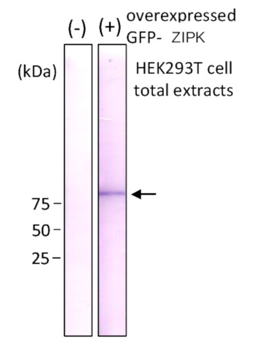
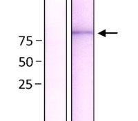
Detection of GFP-ZIPK fusion protein over-expressed in HEK293 cells by Western protein with antibody 1A5. (-) HEK293T cell extract without overexpression (+) GFP-ZIPK protein- overexpressed HEK293T cell extract
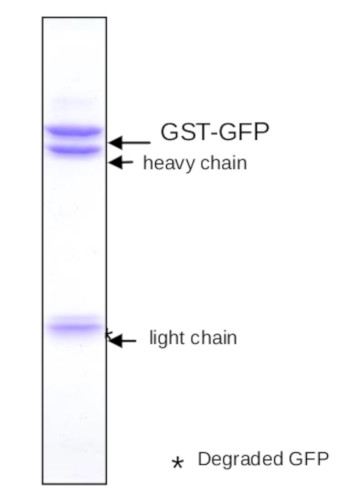
Immunoprecipitation of GST-GFP fusion blotting with antibody 1A5.
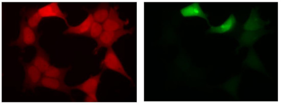
Fluorescent image of COS1 cells with GFP of GST-ZIPK fusion protein expressed in HEK293T cells (Right) and the same cells were immunostained by using anti-GFP antibody 1A5, followed by Texas Red-conjugated anti-rat IgG (Left). Note that fluorescence by the immunofluorescent staining using 1A5 antibody is much stronger than fluorescence due to GFP (right panel). - Additional Information
-
Additional information: Antibody detects GFP and EGFP - Background
-
Background: GFP (Green fluorescent protein) was originally identified in photo organs on jellyfish Aequorea victoria. It is a naturally fluorescent protein which emits green light at a maximum wavelength of 509 nm when excited by blue or UV light. It is extensively used in laboratory as a reporter molecule to label and study cellular and subcellular proteins in living cells using a wide range of applications. Antibodies to GFP protein are used in immunoblotting and ELISA. GFP protein has molecular weight of 27 kDa.
- Product Citations
-
Selected references: Li et al. (2026). Receptor-like-kinase-interacting protein TOW stabilizes PIN transporters for auxin canalization. Curr Biol . 2026 Mar 13:S0960-9822(26)00174-0. doi: 10.1016/j.cub.2026.02.023.
Maehara et al. (2015). issue-specific expression of histone H3 variants diversified after species separation. Epigenetics Chromatin. 2015 Sep 17;8:35. doi: 10.1186/s13072-015-0027-3. (ChIP)
Okazaki et al. (2012). Nuclear localization signal in a cancer-related transcriptional regulator protein NAC1. Carcinogenesis. 2012 Oct;33(10):1854-62.doi: 10.1093/carcin/bgs193. (Immunoprecipiation) - Protocols
- Antibody protocols
- Reviews:
-
This product doesn't have any reviews.
Related products

AS15 2998 | Clonality: Polyclonal | Host: Chicken | Reactivity: native and recombinant GFP
AS10 947 |Clonality: Polyclonal | Host: Donkey | Reactivity: Rat IgG (H&L)