1

Anti-HA (rabbit polyclonal)
AS12 2220 | Clonality: Polyclonal | Host: Rabbit | Reactivity: Human influenza hemagglutinin (HA) tagged proteins (strong cross reactivity between 25 -20 kDa in Arabidopsis thaliana wildtype)
- Product Info
-
Immunogen: KLH-conjugated synthetic peptide: YPYDVPDYA
Host: Rabbit Clonality: Polyclonal Purity: affinity purified serum in PBS, pH 7.4. Format: Lyophilized Quantity: 100 µg Reconstitution: For reconstitution add 100 µl of sterile water Storage: Store lyophilized/reconstituted at -20°C; once reconstituted make aliquots to avoid repeated freeze-thaw cycles. Please remember to spin the tubes briefly prior to opening them to avoid any losses that might occur from material adhering to the cap or sides of the tube. Tested applications: Western blot (WB), immunoprecipitation (IP) Recommended dilution: 1 : 5000 (WB) Expected | apparent MW: Depends on the MW of the protein which is HA-tagged
- Reactivity
-
Confirmed reactivity: HA-tag Not reactive in: No confirmed exceptions from predicted reactivity are currently known - Application Examples
-

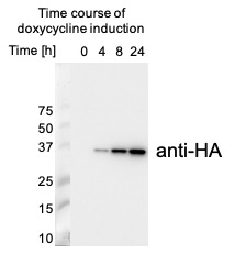
10 µg of total protein extracted freshly from mutant HEK293 cells with RIPA buffer and denatured at 65°C for 5 min were separated on 15% SDS-PAGE and blotted overnight to PVDF (pore size of 0.4 um), using wet transfer. Blot was blocked with 5% milk or 2 h at RT and then incubated in the primary antibody at a dilution of 1: 5 000 in a rotator for 2.5 h in 5% milk. The membrane was then placed in a clean container and rinsed 3 times for 5 min in TBS-T at RT with agitation. Blot was incubated in Agrisera matching secondary antibody (anti-rabbit IgG horse radish peroxidase conjugated, AS09 602) diluted to 1:20 000 in for 1 h at RT with agitation. The blot was washed as above and developed with Agrisera ECLSuperBright. Exposure time was 30 seconds.
Courtesy Dr. Małgorzata Wessels, Department of Medical Biochemistry and Biophysics, Umeå University, Sweden
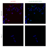
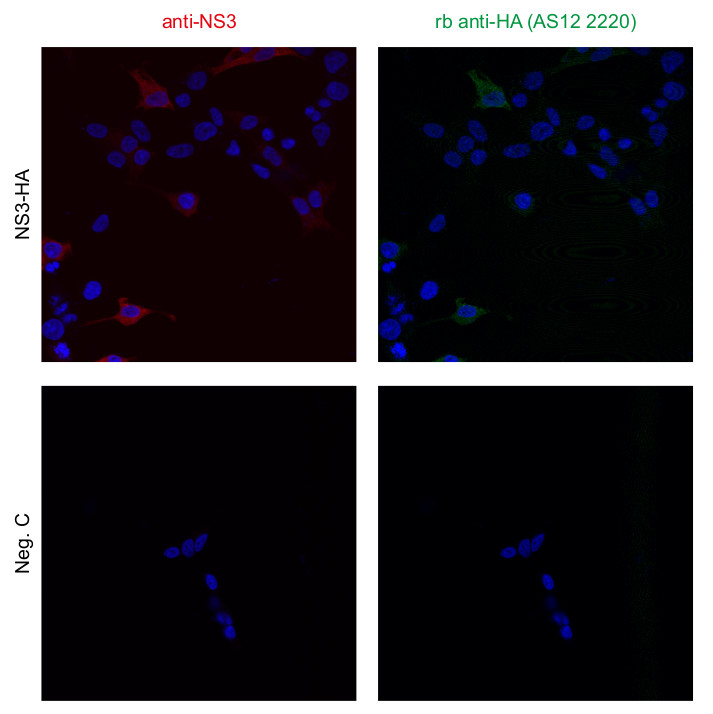
HEK293 cells were transfected with the indicated plasmid (SARS-CoV-2 N myc tagged, PMID: 34799561, TBEV NS3-HA, PMID: 29321318, Rab5 mcherry, Addgene: 4920, GFP-HIS) using genejuice transfection reagent (EMD Millipore) according to the manufacturer’s instructions. After 24 hours of transfection, cells were fixed in 4% formaldehyde and permeabilized in PBS containing 0.5% Triton X-100 and 20 mM glycine. Then, cells were stained with the primary anti-HA epitope tag, rabbit polyclonal antibodies at a concentration of 1 μg/mL for 1 hour at room temperature. Followed by three washes in PBS. Cells were then stained using secondary antibodies, donkey antirabbit Alexa488 (1:500, Thermo Fisher Scientific, a21206 in PBS containing 2% BSA for 1h at RT. Nuclei were stained using DAPI (1 μg/mL). Images were acquired using a Leica SP8 Laser Scanning Confocal Microscope with a 63x oil objective (Leica) and Leica Application Suit X software (LAS X, Leica). Anti-NS3 antibodies are used as a control.
Courtesy of Dr. Anna K Överby, Molecular Infection Medicine Sweden (MIMS),Section of Virology. Department of Clinical Microbiology Umeå University, Sweden
- Additional Information
-
Additional information (application): Important note: there is a strong cross reactivity between 25 -20 kDa in Arabidopsis thaliana wildtype. - Background
-
Background: HA-tag is derived from a human influenza hemagglutinin HA-molecule corresponding to amino acids 96-106 and is used as a general epitope tag in expression vectors. - Product Citations
-
Selected references: Tanigawa et al. (2024). FYVE1/FREE1 is involved in glutamine-responsive TORC1 activation in plants. iScience. 2024 Aug 26;27(9):110814. doi: 10.1016/j.isci.2024.110814.
Hwang et al. (2019) Arabidopsis ABF3 and ABF4 Transcription Factors Act with the NF-YC Complex to Regulate SOC1 Expression and Mediate Drought-Accelerated Flowering. Mol Plant. 2019 Apr 1;12(4):489-505. doi: 10.1016/j.molp.2019.01.002 - Protocols
-
Antibody protocols
- Reviews:
-
This product doesn't have any reviews.


