1

Anti-L13-1 | 60S ribosomal protein L13-1
AS13 2650 | Clonality: Polyclonal | Host: Rabbit | Reactivity: Arabidopsis thaliana, Hordeum vulgare, Nicotiana benthamiana, Zea mays
- Product Info
-
Immunogen: KLH-conjugated synthetic peptide derived from Arabidopsis thaliana UniProt: P41127, TAIR: At3g49010
Host: Rabbit Clonality: Polyclonal Purity: Antigen affinity purified serum in PBS pH 7.4. Format: Lyophilized Quantity: 50 µg Reconstitution: For reconstitution add 50 µl of steril water Storage: Store lyophilized/reconstituted at -20°C; once reconstituted make aliquots to avoid repeated freeze-thaw cycles. Please remember to spin the tubes briefly prior to opening them to avoid any losses that might occur from material adhering to the cap or sides of the tube. Tested applications: Western blot (WB) Recommended dilution: 1 : 2500 (WB) Expected | apparent MW: 23.7 | 29 kDa (Arabidopsis thaliana)
- Reactivity
-
Confirmed reactivity: Arabidopsis thaliana, Hordeum vulgare, Nicotiana benthamiana, Zea mays Predicted reactivity: Brassica napus, Chlamydomonas reinhardii, Nicotiana tabacum, Oryza sativa, Picea excelsa, Populus balsamifera, Sorghum bicolor, Ricinus communis, Vitis vinifera
Species of your interest not listed? Contact usNot reactive in: Diatoms - Application Examples
-
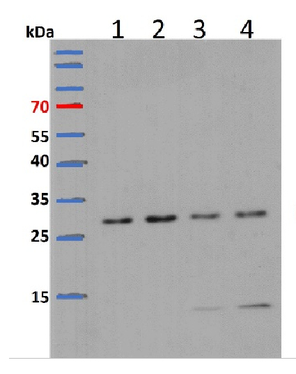
Samples:
1 - 15 ug of Arabidopsis thaliana whole seedling extract
2 - 15 ug of Arabidopsis thaliana whole seedling extract
3 - 15 ug of Arabidopsis thaliana whole seedling treated with Drug-1
4 - 15 ug of Arabidopsis thaliana whole seedling treated with Drug-2
15 µg/well of total protein extracted freshly from Arabidopsis thaliana 8-day-old seedlings extracted with buffer containing 8 M Urea. Samples were separated in the RT on 12 % SDS-PAGE and blotted for 2 h to PVDF (pore size of 0.2 um), using: wet transfer in the cold. Blot was blocked with 10 % milk for 1h/RT with agitation. Blot was incubated in the primary antibody at a dilution of 1: 3000 for 1h/RT in PBS with agitation. The antibody solution was decanted, and the blot was rinsed briefly twice, then washed once for 15 min and 3 times for 5 min in PBS at RT with agitation. Blot was incubated in matching secondary antibody (anti-rabbit IgG horse radish peroxidase conjugated) diluted to 1: 5000 in for 1 h/RT with agitation. The blot was washed as above and developed with a chemiluminescent detection reagent. Exposure time was 1 minute.Courtesy of Dr. Kuo-En Chen, Department of Biology, Washington University in St. Louis, USA
- Additional Information
-
Additional information (application): Protein or membrane sample should be treated at 70°C for 10 min before loading on the gel - Background
-
Background: L13 protein is localized in the cytoplasm. It belongs to the ribosomal protein L13e family. Alternative name: protein BBC1 homolog.
- Product Citations
-
Selected references: González-Fuente et al. (2026). Bacteria use P-body condensates to attenuate host translation during infection. Sci Adv . 2026 Apr 24;12(17):eaec4477. doi: 10.1126/sciadv.aec4477.
Zhong et al. (2026). CDS-localized m6A drives co-translational RNA decay to relieve biotic and abiotic endoplasmic reticulum stresses. Nat Plants . 2026 May 8. doi: 10.1038/s41477-026-02299-4.
Wang et al. (2024). Maize Dek51 encodes a DEAD-box RNA helicase essential for pre-rRNA processing and seed development. Cell Rep. 2024 Sep 24;43(9):114673. doi: 10.1016/j.celrep.2024.114673.
Lee et al. (2023). The Phytophthora nucleolar effector Pi23226 targets host ribosome biogenesis to induce necrotrophic cell death. Plant Commun. 100606. doi: 10.1016/j.xplc.2023.100606.
Pereira Firmino et al. (2020). Separation and Paired Proteome Profiling of Plant Chloroplast and Cytoplasmic Ribosomes. Plants (Basel) . 2020 Jul 14;9(7):892.doi: 10.3390/plants9070892.
Shinozaki et al. (2020). Autophagy Increases Zinc Bioavailability to Avoid Light-Mediated ROS Production under Zn Deficiency. Plant Physiol. 2020 Jan 15. pii: pp.01522.2019. doi: 10.1104/pp.19.01522.
Li (2019). The Isolation of Total and Membrane-Bound Polysomes from Arabidopsis and the Detection of Their Associated AGO1 and sRNAs. Methods Mol Biol. 2019;1932:317-333. doi: 10.1007/978-1-4939-9042-9_23.
You et al. (2019). FIERY1 promotes microRNA accumulation by suppressing rRNA-derived small interfering RNAs in Arabidopsis. Nat Commun. 2019 Sep 27;10(1):4424. doi: 10.1038/s41467-019-12379-z.
de Francisco Amorim et al. (2018). The U1 snRNP Subunit LUC7 Modulates Plant Development and Stress Responses via Regulation of Alternative Splicing. Plant Cell. 2018 Nov;30(11):2838-2854. doi: 10.1105/tpc.18.00244. Epub 2018 Oct 11.
Beine-Golovchuk et al. (2018). Plant Temperature Acclimation and Growth Rely on Cytosolic Ribosome Biogenesis Factor Homologs. Plant Physiol. 2018 Mar;176(3):2251-2276. doi: 10.1104/pp.17.01448. Epub 2018 Jan 30.
Wang et al. (2016). Comprehensive proteomic analysis of developing protein bodies in maize (Zea mays) endosperm provides novel insights into its biogenesis. J Exp Bot. 2016 Dec;67(22):6323-6335. Epub 2016 Oct 27. - Protocols
-
Agrisera Western Blot protocol and video tutorials
Protocols to work with plant and algal protein extracts
Oxygenic photosynthesis poster by prof. Govindjee and Dr. Shevela
Z-scheme of photosynthetic electron transport by prof. Govindjee and Dr. Björn and Dr. Shevela - Reviews:
-
J ZHU | 2024-01-24The antibody works well in Arabidopsis!
Related products

AS09 602 | Clonality: Polyclonal | Host: Goat | Reactivity: Rabbit IgG (H&L)

AS09 607 | Clonality: Polyclonal Host: Goat Reactivity: Rabbit IgG (H&L)
AS11 1738 | Clonality: Polyclonal | Host: Rabbit | Reactivity: cyanobacteria