1
Anti-PIF3 | Phytochrome interacting factor 3 (goat antibody)
AS16 3954 | Clonality: Polyclonal | Host: Goat | Reactivity: Arabidopsis thaliana
- Data sheet
- Product Info
-
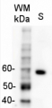
Immunogen: KLH-conjugated peptide, derived from Arabidopsis thaliana PIF3, UniProt: O80536, TAIR: AT1G09530 Host: Goat Clonality: Polyclonal Purity: Immunogen affinity purified serum in PBS pH 7.4. Format: Lyophilized Quantity: 50 µg Reconstitution: For reconstitution add 50 µl of sterile water Storage: Store lyophilized/reconstituted at -20°C; once reconstituted make aliquots to avoid repeated freeze-thaw cycles,Please remember to spin the tubes briefly prior to opening them to avoid any losses that might occur from material adhering to the cap or sides of the tube. Tested applications: Western blot (WB) Recommended dilution: 1 : 1000 (WB) Expected | apparent MW: 57 | ca. 65 kDa - Reactivity
-
Confirmed reactivity: Arabidopsis thaliana Predicted reactivity: Cardamine hirsuta
Species of your interest not listed? Contact usNot reactive in: Citrus sp., Daucus carota, Marchantia polymorpha, Nicotiana attenuata, Solanum lycopersicum, Triticum aestivum, Vitis vinifera - Application Examples
-
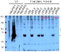
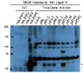
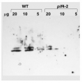
Application example 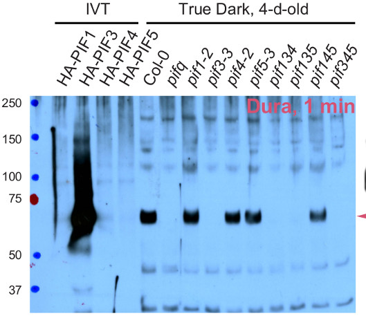
Total protein from Arabidopsis thaliana (10-20 µg) of 4-d-old dark grown Col-0, pif single, triple and quadruple mutants were extracted with the buffer described in Qiu et al. 2015 and denatured at 95°C for 5 min. were separated on 8 % Bis-Tris SDS-PAGE, and subsequently blotted to Nitrocellulose membrane using wet tank transfer. Blots were blocked with TBS containing 2% non-fat milk at room temperature for 1h. Primary anti-PIF3 antibodies were used in dilution of 1:1000 to TBS containing 2% milk. The incubation time was 14-20 hours at 4°C. After washing 4x5 min. with TBST at room temperature, the blots were incubated with HRP-conjugated secondary antibodies (1:5000) at room temperature for 1h. The blots were washed 4x10 min with TBST again and the signal was detected using the SuperSignal West Dura Extended Duration Substrate (ThermoFisher Scientific).
Courtesy od Dr. Yongjian Qiu, University of California-Riverside, USAApplication examples: 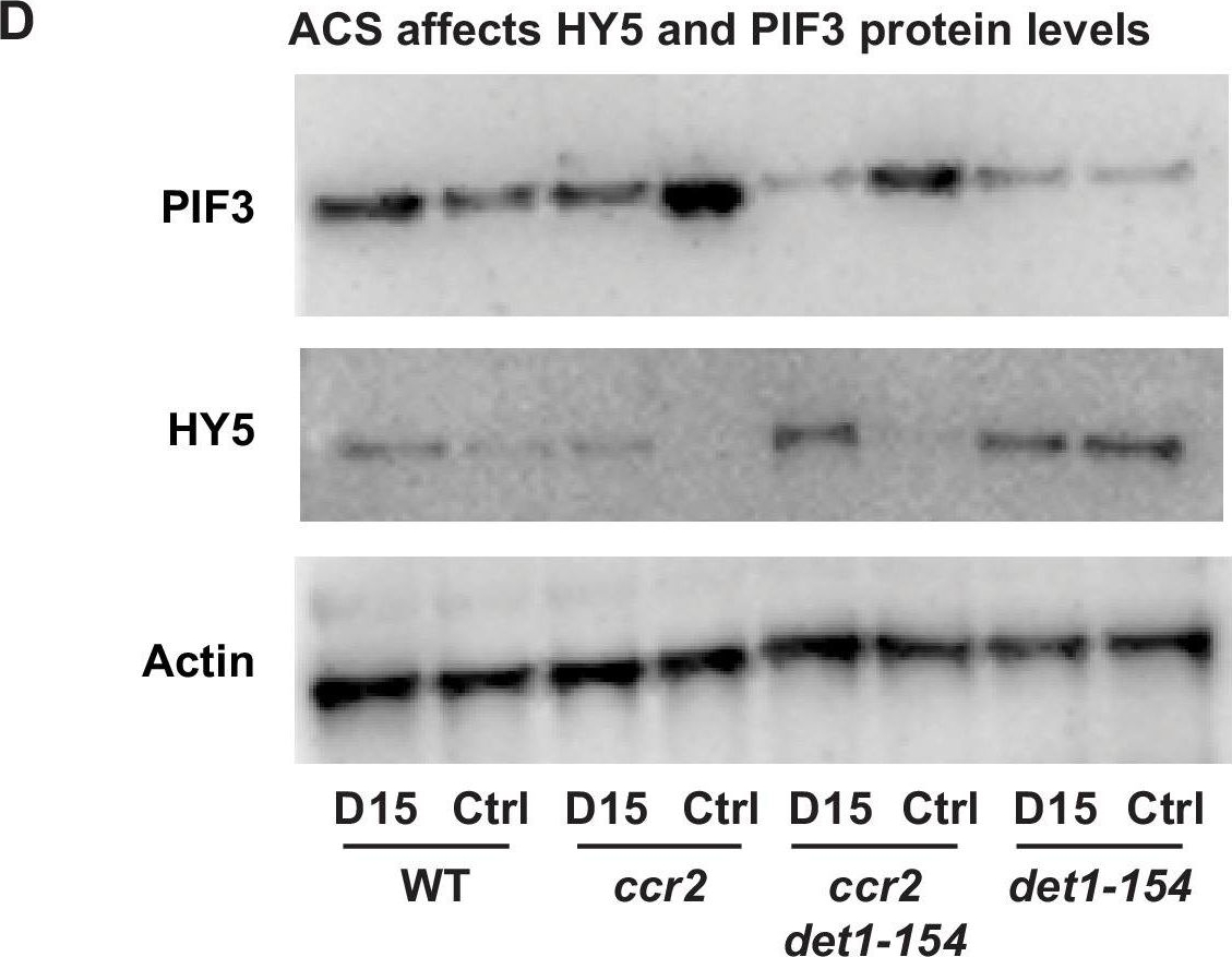
Reactant: Arabidopsis thaliana (Thale cress)
Application: Western Blotting
Pudmed ID: 32003746
Journal: Elife
Figure Number: 7D
Published Date: 2020-01-31
First Author: Cazzonelli, C. I., Hou, X., et al.
Impact Factor: 7.448
Open PublicationChemical inhibition of CCD activity revealed how a ccr2 generated apocarotenoid signal transcriptionally up-regulates POR and PIF3 in parallel to det1-154 during skotomorphogenesis.(A) Transcript levels of PORA, PIF3 and HY5 in WT, ccr2, ccr2 det1-154 and det1-154 etiolated seedlings growing on MS media (+ /- D15). Statistical analysis denoted as a star was performed by a pair-wise t-test (p<0.05). Error bars represent standard error of means. (B), (C) and (D) Representative western blot images showing POR, DET1, PIF3 and HY5 protein levels, respectively. Proteins were extracted from WT, ccr2 and ccr2 det1-154 etiolated seedlings grown on MS media without (control; Ctrl) or with the chemical inhibitor of CCD activity (D15). The membrane was re-probed using anti-Actin antibody as an internal loading control. Lattice-like symbol below POR western (B), represents formation of a PLB in etiolated cotyledons from that genotype and treatment. (E) Model describing how a cis-carotene derived cleavage product, ACS, regulates POR, HY5, PIF3 and PLB formation during skotomorphogenesis. DET1 maintains skotomorphogenesis by post-transcriptionally maintaining a higher and lower PIF3 and HY5 protein levels, respectively. HY5 promotes and PIF3 represses PhANG expression. det1 mutants trigger photomorphogenesis in that they lack POR mRNA transcripts, protein and a PLB. ccr2 generates ACS that enhances POR mRNA transcript and protein levels that enable PLB formation in det1-154. det1-154 restores PLB formation in ccr2 by blocking a signalling pathway acting independent of POR.The DET1-154 peptide is smaller in det1-154 mutant genotypes.(A) Representative western blot image showing the reduced DET1 peptide size in ccr2 det1-154 and det1-154 (59 kDa) compared to WT and ccr2 (62 kDa). Gel electrophoresis of the gel membrane from Figure 7C (electrophoresis for 38 min at 165 volt) was extended for 120 min at 100 volts to resolve the 3 kDa difference in DET1-154 protein size. Under these conditions, the 37 kDa ACTIN peptide and pre-stained ladder were not detected on the membrane. Proteins were extracted from WT, ccr2 and ccr2 det1-154 etiolated seedlings grown on MS media without (control; Ctrl) or with the chemical inhibitor of CCD activity (D15).
Reactant: Arabidopsis thaliana (Thale cress)
Application: Western Blotting
Pudmed ID: 32003746
Journal: Elife
Figure Number: 8B
Published Date: 2020-01-31
First Author: Cazzonelli, C. I., Hou, X., et al.
Impact Factor: 7.448
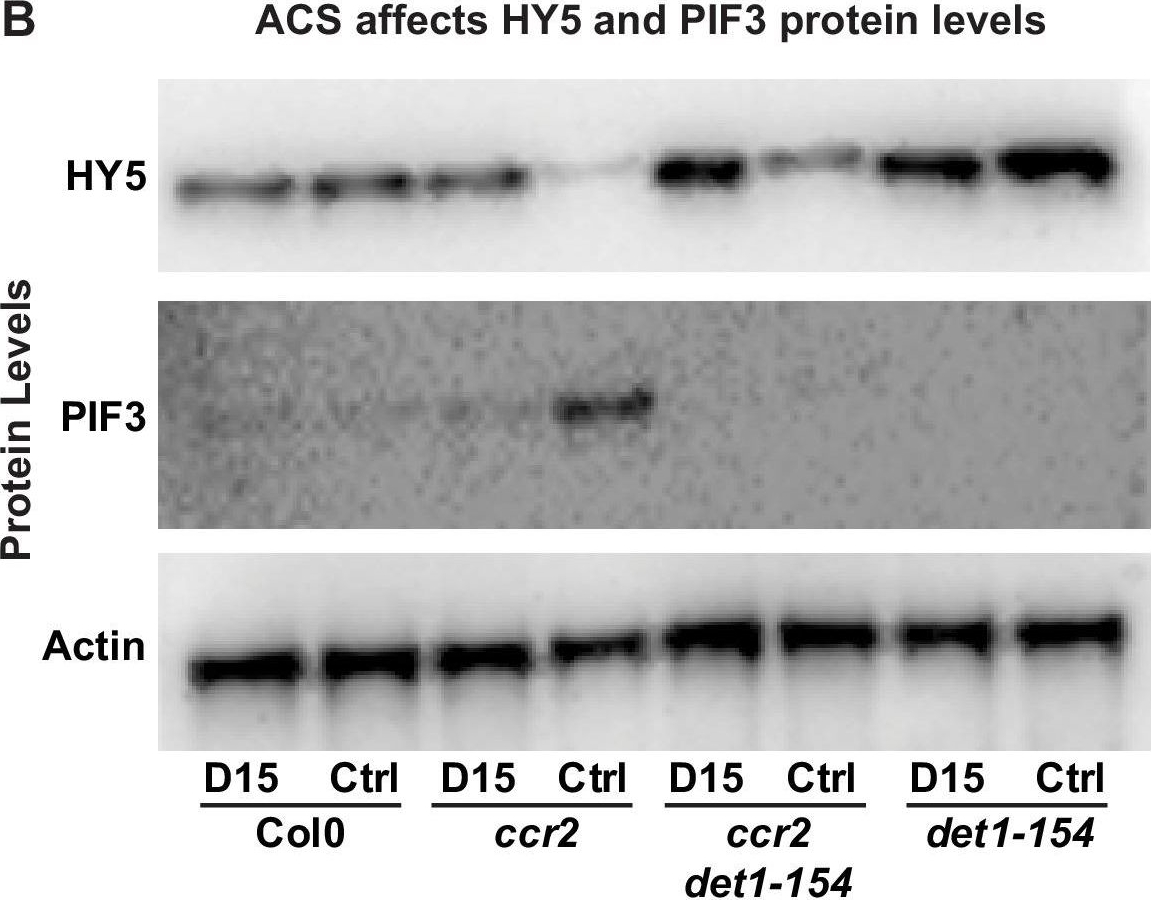
Open PublicationChemical inhibition of CCD activity revealed how a ccr2 generated apocarotenoid signal transcriptionally represses HY5 and LHCB2 expression during photomorphogenesis.(A) Transcript levels of PIF3 and HY5 in WT, ccr2, ccr2 det1-154 and det1-154 de-etiolated seedlings growing on MS media + /- D15. (B) Representative western blot images showing PIF3 and HY5 protein levels in WT, ccr2, ccr2 det1-154 and det1-154 de-etiolated seedlings growing on MS media + /- D15. The membrane was re-probed using anti-Actin antibody as an internal loading control. (C) Protein and transcript levels of LHCB2 expression in WT and ccr2 de-etiolated seedlings growing on MS media + /- D15. (D) Model showing how ACS regulates HY5 and LHCB2 expression in ccr2. Images of seedlings represent are cotyledons are coloured green or yellow to reflect the delay in chlorophyll biosynthesis induced by ACS as evidenced in Figure 6c. De-etiolation of seedlings was performed by transferring 4-d-old etiolated seedlings to continuous light for 3 d to induce photomorphogenesis. Statistical analysis denoted as a star was performed by pair-wise t-test (p<0.05). Error bars represent standard error of means. Ctrl; Control; Ctrl, D15; chemical inhibitor of CCD activity.
- Additional Information
-
Additional information: Please, do not re-use PIF3 antibody solution after first incubation with your membrane as most of antibody will bind in this step and next result will not be reproducable Additional information (application): PIF3 protein runs at higher MW than expected, as observed previously (Al-Sady et al. 2006). PIF proteins are not that stable, therefore special precautions should be taken during extraction and whole procedure should be performed in as little light as possible (light green light).
- Background
-
Background: PIF3 (Phytochrome interacting factor 3) is a transcription factor which acts positively in the phytochrome signaling pathway. It activates transcription by binding to the G box. Subcellular localization is nucleus. Alternative names:Basic helix-loop-helix protein 8, AtbHLH8, bHLH8, Phytochrome-associated protein 3, Phytochrome-interacting factor 3, Transcription factor EN 100, EN100, bHLH transcription factor bHLH008, PAP3, PHYTOCHROME-ASSOCIATED PROTEIN 3, POC1, PHOTOCURRENT 1,ABI1, ABA INSENSITIVE 1, AtABI1. - Product Citations
-
Selected references: Sinclair et al. (2017) Etiolated Seedling Development Requires Repression of Photomorphogenesis by a Small Cell-Wall-Derived Dark Signal. Curr Biol. 2017 Nov 20;27(22):3403-3418.e7. doi: 10.1016/j.cub.2017.09.063. - Protocols
-
Agrisera Western Blot protocol and video tutorials
Protocols to work with plant and algal protein extracts
Agrisera Educational Posters Collection - Reviews:
-
Aashish Ranjan | 2021-09-30Worked great for both western and ChIP experiments with Arabidopsis leaves. We used 1:1000 dilution for western and 5ul for ChIP experiments.
Related products
AS12 2112 | Clonality: Polyclonal | Host: Rabbit | Reactivity: Arabidopsis thaliana
AS16 3157 | Clonality: Polyclonal | Host: Rabbit | Reactivity: Arabidopsis thaliana