1
Anti-Tubulin beta chain
AS10 681 | Clonality: Polyclonal | Host: Rabbit | Reactivity:Arabidopsis thaliana, Medicago sativa
- Product Info
-
Immunogen: KLH-conjugated peptide derived from available tubulin beta chain sequences (located at a surface loop) including all Arabidopsis thaliana tubulin beta-2/beta-3 chain P29512, beta-4 chain P24636(At5g44340), beta-5 chain P29513(At1g20010), beta-6 chain P29514(At5g12250), beta-7 chain P29515(At2g29550), beta-8 chain P29516(At5g23860), beta-9 chain P29517(At4g20890).
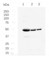
Host: Rabbit Clonality: Polyclonal Purity: Immunogen affinity purified serum in PBS pH 7.4. Format: Lyophilized Quantity: 100 µg Reconstitution: For reconstitution add 100 µl of sterile water Storage: Store lyophilized/reconstituted at -20°C; once reconstituted make aliquots to avoid repeated freeze-thaw cycles. Please remember to spin the tubes briefly prior to opening them to avoid any losses that might occur from material adhering to the cap or sides of the tube. Tested applications: immunofluorescence (IF), Western blot (WB) Recommended dilution: 1 : 1000 (IF), 1 : 500 (WB) Expected | apparent MW: 49 | 49 kDa (Arabidopsis thaliana)
- Reactivity
-
Confirmed reactivity: Arabidopsis thaliana (including suspension cells), Chlamydomonas reinhardii, Medicago sativa Predicted reactivity: Brassica napus, Chlamydomonas reinhardii, Glycine max, Hordeum vulgare, Micromonoas pusilla, Nicotiana attenuata, Oryza sativa, Ostreococcus lucimarinus, Picea sitcHensis, Pisum sativum, Physcomitrium patens, Populus trichocarpa, Pythium ultimum, Saccharomyces cerevisiae, Solanum tuberosum, Sorghum bicolor, Ricinus communis, Zea mays, Vigna radiata, Vitis vinifera
Species of your interest not listed? Contact usNot reactive in: Allium sativum - Application Examples
-
Application example

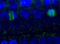
Tubulin beta localization in Arabidopsis thaliana epidermis cells. Tubulin beta localized to division plates visualized in green, nucleus with DAPI in blue. Plant material has been fixed in para-formaldehyde for 30 minutes. Tissue cleaning has been performed before immunolocalization. Primary antibodies: Agrisera anti-tubulin beta priamry antibody in dilution 1: 1000 and Agrisera goat anti-rabbit IgG DyLight® 488, secondary antibody in dilution 1: 2000. Scale bar – 10 µm.

(A) The 3 days old suspension cells of Arabidopsis thaliana have been fixed for 25 minutes with 2% FA, cell wall was partaillly digested with 0.2% Dricelase in MES buffer (pH 5.1) for 30 min at 37°C and the cells were dried on coated slide to omit PM extraction. Following by incubation for 45 minutes with Agrisera anti-tubulin beta chain primary antibody in a dilution of 1: 1000, washing once and 45 min incubation with a secondary goat anti-rabbit Alexa 555 antibody, in a dilution of 1:1000 dilution (Invitrogen). Cells were counterstained witn DAPI and visialized under epi/fluiresecnet microscope (B).
Courtesy Dr. Taras Pasternak, Freiburg University, Germany
Application examples: 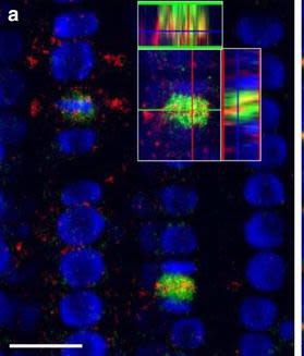
Reactant: Arabidopsis thaliana (Thale cress)
Application: Immunohistochemistry-immunofluorescence
Pudmed ID: 26516341
Journal: Plant Methods
Figure Number: 7A
Published Date: 2015-10-31
First Author: Pasternak, T., Tietz, O., et al.
Impact Factor: 5.139
Open Publication3D reconstruction of Arabidopsis root epidermis cells undergoing telophase: co-localization of ?-Tubulin (TUB) and PIN1 in division plates. Four days old Arabidopsis seedlings were fixed for 30 min in formaldehyde. a Anti-PIN1 mouse monoclonal primary antibody (10A7) diluted 1:50 plus ALEXA Fluor ® 555 anti-mouse as secondary antibody diluted 1:800 (shown in green color) and anti-TUB (AS10 681) rabbit primary antibody diluted 1:600 plus Goat anti-rabbit IgG (H&L), DyLight® 488 Conjugate (AS09 633) diluted in 1:3000 as secondary antibody (shown in red color) were used. b Anti-PIN2 Guinea pig polyclonal primary antibody (clone A193) dilution 1:800 plus ALEXA Fluor ® 555 anti-Guinea pig as the secondary antibody dilution 1:800 (show in green color) and anti-TUB (Agrisera, AS10 681) rabbit primary antibody diluted 1:600 plus Goat anti-rabbit IgG (H&L), DyLight® 488 Conjugate (AS09 633) as secondary antibody diluted in 1:3000 (shown in red color) were used. Co-staining with DAPI visualizes nucleus (blue). Scale bar 20 µm. The Insertion in a shows an ortho-view of dividing cells
Reactant: Arabidopsis thaliana (Thale cress)
Application: Immunohistochemistry-immunofluorescence
Pudmed ID: 26516341
Journal: Plant Methods
Figure Number: 7b
Published Date: 2015-10-31
First Author: Pasternak, T., Tietz, O., et al.
Impact Factor: 5.139
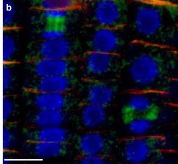
Open Publication3D reconstruction of Arabidopsis root epidermis cells undergoing telophase: co-localization of ?-Tubulin (TUB) and PIN1 in division plates. Four days old Arabidopsis seedlings were fixed for 30 min in formaldehyde. a Anti-PIN1 mouse monoclonal primary antibody (10A7) diluted 1:50 plus ALEXA Fluor ® 555 anti-mouse as secondary antibody diluted 1:800 (shown in green color) and anti-TUB (AS10 681) rabbit primary antibody diluted 1:600 plus Goat anti-rabbit IgG (H&L), DyLight® 488 Conjugate (AS09 633) diluted in 1:3000 as secondary antibody (shown in red color) were used. b Anti-PIN2 Guinea pig polyclonal primary antibody (clone A193) dilution 1:800 plus ALEXA Fluor ® 555 anti-Guinea pig as the secondary antibody dilution 1:800 (show in green color) and anti-TUB (Agrisera, AS10 681) rabbit primary antibody diluted 1:600 plus Goat anti-rabbit IgG (H&L), DyLight® 488 Conjugate (AS09 633) as secondary antibody diluted in 1:3000 (shown in red color) were used. Co-staining with DAPI visualizes nucleus (blue). Scale bar 20 µm. The Insertion in a shows an ortho-view of dividing cells
- Additional Information
-
Additional information: Please, note that tubulin beta is only detected in actively dividing meristematic cells (see immunolocalization image below). Therefore to allow detection on a western blot, analyzed material must contain enough meristematic cells with dividing activity.
Signal in a western blot application in Chlamydomonas reinhardtii is obtained with a load of 100 µg/well and a dilution of 1: 500.Additional information (application): Metal induced stress affected the expression of tubulin, and that therefore, this protein cannot be used as a loading control under that type of conditions. More information can be found here. - Background
-
Background: Tubulin beta (TUB) together with alpha tubulin is making up microtubules.
- Product Citations
-
Selected references: Gong et al. (2024).HYPK controls stability and catalytic activity of the N-terminal acetyltransferase A in Arabidopsis thaliana. Cell Rep. 2024 Feb 15;43(2):113768.doi: 10.1016/j.celrep.2024.113768.
Durian et al. (2019). PROTEIN PHOSPHATASE 2A-B' gamma controls Botrytis cinerea resistance and developmental leaf senescence. Plant Physiol. 2019 Oct 28. pii: pp.00893.2019. doi: 10.1104/pp.19.00893.
Wang et al. (2017). The inhibition of protein translation mediated by AtGCN1 is essential for cold tolerance in Arabidopsis thaliana. Plant Cell Environ. 2017 Jan;40(1):56-68. doi: 10.1111/pce.12826.
Heinnickel et al. (2016). Tetratricopeptide repeat protein protects photosystem I from oxidative disruption during assembly. Proc Natl Acad Sci U S A. 2016 Mar 8;113(10):2774-9. doi: 10.1073/pnas.1524040113 - Protocols
-
Agrisera Western Blot protocol and video tutorials
Protocols to work with plant and algal protein extracts - Reviews:
-
This product doesn't have any reviews.
Related products

AS09 602 | Clonality: Polyclonal | Host: Goat | Reactivity: Rabbit IgG (H&L)

AS09 607 | Clonality: Polyclonal Host: Goat Reactivity: Rabbit IgG (H&L)
AS10 680 | Clonality: Polyclonal | Host: Rabbit | Reactivity: A. thaliana, C. reinhardii, E. gracilis, H. vulgare, M. crystallinum, O. sativa, Setaria italica, Zea mays