2
Anti-BiP | Lumenal-binding protein (goat antibody)
AS09 615 | Clonality: Polyclonal | Host: Goat | Reactivity: Arabidopsis thaliana, Spinacia oleracea
- Product Info
-
Immunogen: KLH-conjugated synthetic peptide derived from Arabidopsis thaliana BiP proteins: BiP1 UniProt:Q9LKR3, TAIR: At5g28540, BiP2 UniProt: F4K007, TAIR: At5g42020, BiP3 UniProt:Q8H1B3 ,TAIR:At1g09080 Host: Goat Clonality: Polyclonal Purity: Immunogen affinity purified serum in PBS pH 7.4. Format: Lyophilized Quantity: 100 µg Reconstitution: For reconstitution add 100 µl of sterile water Storage: Store lyophilized/reconstituted at -20°C; once reconstituted make aliquots to avoid repeated freeze-thaw cycles. Please remember to spin the tubes briefly prior to opening them to avoid any losses that might occur from material adhering to the cap or sides of the tube. Tested applications: Western blot (WB) Recommended dilution: 1 : 2000 (WB) Expected | apparent MW: 73.5 | 80 kDa - Reactivity
-
Confirmed reactivity: Arabidopsis thaliana, Hordeum vulgare, Spinacia oleracea, Zea mays Predicted reactivity: Hordeum vulgare, Nicotiana tabacum, Oryza sativa, Picea sitchensis, Populus trichocarpa, Physcomitrium patens, Spinacia oleracea, Zea mays
Species of your interest not listed? Contact usNot reactive in: No confirmed exceptions from predicted reactivity are currently known - Application Examples
-
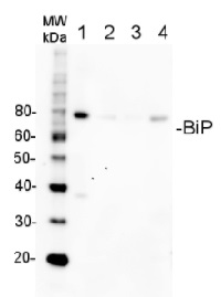
Application example

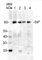
5 µg of total protein from A.thaliana (1), H. vulgare (2), Z.mays (3), S. oleracea (4), extracted with Agrisera PEB extraction buffer (AS08 300) were separated on 4-12% SDS-PAGE and blotted 1h to PVDF. Blots were blocked immediately following transfer in for 1h at room temperature with agitation. Blots were incubated in the primary antibody at a dilution of 1: 10 000 for 1h at room temperature with agitation. The antibody solution was decanted and the blot was rinsed briefly twice, then washed once for 15 min and 3 times for 5 min in TBS-T at room temperature with agitation. Blots were incubated in secondary antibody (anti-goat IgG horse radish peroxidase conjugated, from Agrisera AS09 605) diluted to 1:50 000 for 1h at room temperature with agitation. The blots were washed as above and developed for 5 min with ECL detection reagent according to the manufacturers instructions. Exposure time was 5 seconds.
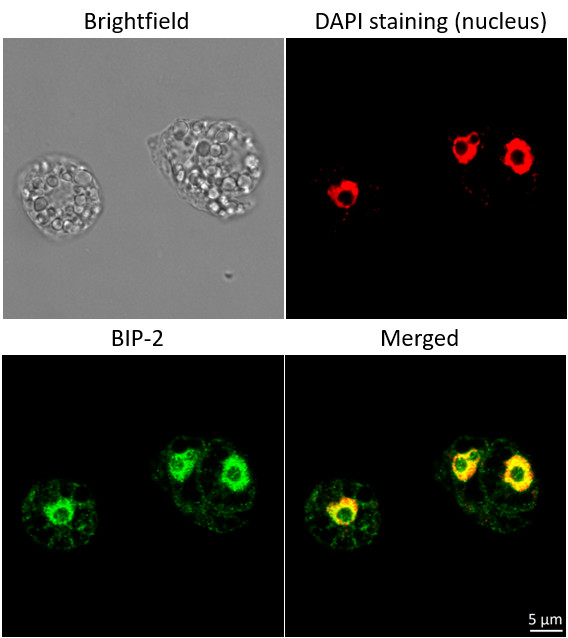
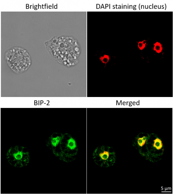
Immunofluorescent localization of BiP in suspension cultures of Arabidopsis thaliana (Landsberg erecta cv. MM1) using goat anti-BiP polyclonal antibodies (AS09 615) and donkey anti-goat IgG DyLight®488 conjugated secondary antibodies (AS10 1116, Agrisera).Material: suspension cultures of Arabidopsis thaliana ecotype Landsberg erecta cv. MM1)
Fixation: Packed cell volume to fixer ratio: 250 µl : 5ml
Fixer composition and buffer: 4% (w/v) paraformaldehyde (freshly prepared as 8% stock and 0.2 µm filtered) 0.01% (v/v) Triton-X100 in Phosphate Buffered Saline (PBS), pH 7.4 (2x stock, 0.2 µm filtered)
Container and method: in 6 cm Petri dish, gentle shaking at room temperature (RT)
Duration: 25 min.
Hydrophilization: No
Cell wall digestion: Yes
Packed cell volume to enzyme ratio: 100 ul : 2ml Enzyme composition: 1% Cellulase (chromatically purified, powder, Worthington) 1% Pectinase (protease free, liquid, Sigma) Buffer: 0.5% (w/v) MES buffer, pH 5.6 Container and method: in 2 ml microfuge tube by rolling at room temperature (RT) Duration: 30 min.
Membrane permeabilization: Triton-X100 (0.5%), 10 min/RT
Antigen retrieval: No
Blocking buffer: Fish gelatin (5% v/v)
Washing buffer: PBS
Primary antibody dilution and incubation time: 1:400, ON/4ºC
Secondary antibody: donkey anti-goat IgG DyLight®488 conjugated secondary antibodies (AS10 1116, Agrisera), 1: 600, 1h/RT
Co-staining of the nucleus (DAPI): Yes
Cell wall and nucleus staining: 100 ng/ml DAPICourtesy of Dr. Ferhan Ayaydin, Hungarian Centre of Excellence for Molecular Medicine (HCEMM), Szeged, Hungary
- Additional Information
-
Additional information (application): Protein or membrane sample should be treated at 70°C for 10 min before loading on the gel.
Antibody has a reduced reactivity to monocots in western blot.
- Background
-
Background: BiP2 (Binding immunoglobulin protein) is localized in endoplasmic reticulum lumen (ER) and plays a role in protein assembly inside ER. BiP protein is abundant under all growth conditions but its synthesis can increase under conditions that lead to the accumulation of unfolded polypeptides in endoplasmic reticulum (ER). Alternative name: AtBP2
- Product Citations
-
Selected references: Narusaka et al (2016). Leucine zipper motif in RRS1 is crucial for the regulation of Arabidopsis dual resistance protein complex RPS4/RRS1. Sci Rep. 2016 Jan 11;6:18702. doi: 10.1038/srep18702. - Protocols
-
Agrisera Western Blot protocol and video tutorials
Protocols to work with plant and algal protein extracts
Oxygenic photosynthesis poster by prof. Govindjee and Dr. Shevela
Z-scheme of photosynthetic electron transport by prof. Govindjee and Dr. Björn and Dr. ShevelaMethod of isolation of plant ER
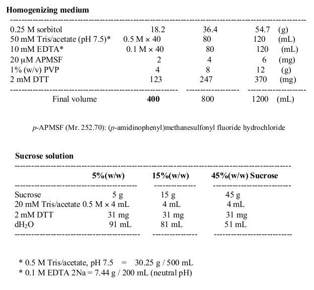
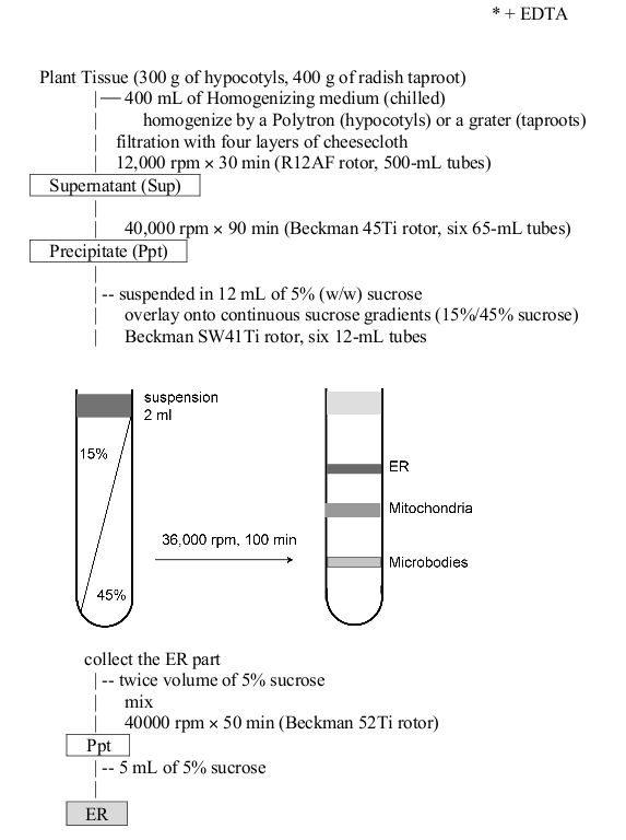
Courtesy of Dr. Masayoshi Maeshima, Laboratory of Cell Dynamics, Graduate School of Bioagricultural Sciences Nagoya University Nagoya, Japan
- Reviews:
-
This product doesn't have any reviews.
Related products
AS09 614 | Clonality: Polyclonal | Host: Hen | Reactivity: Arabidopsis thaliana, Hordeum vulgare, Spinacia oleracea, Zea mays
Limited stock
AS09 630 | Clonality: Polyclonal | Host: Rabbit | Reactivity: Goat IgG (H&L)