| Can the exact same extraction and Western blot protocols be applied to detect a given target protein in samples from different species? As you will see in the example to the right, the quality and intensity of the PetA band in each analyzed species is different. The following samples were extracted: 1. Picea abies total proteins extract obtained from needles using 10% TCA/acetone, pellet was dissolved in 8M Urea, 40 mM Tris-HCl, pH 6.8, 0,1 mM EDTA, 1% SDS 2. Pinus sylvestris total proteins extract obtained from needles using 10% TCA/acetone, pellet was dissolved in 8M Urea, 40 mM Tris-HCl, pH 6.8, 0,1 mM EDTA, 1% SDS 3. Lupinus angustifolius thylakoid membranes 4. Solanum tuberosum thylakoid membranes 5. Synechococcus elongatus PCC7942 thylakoid membranes 6. Pisum sativum, thylakoid membranes Samples from conifers required another type of extraction compared to samples from other plant species. In the case of cyanobacterial sample, a higher protein load per well and longer incubation time of the primary antibody is required. This result clearly shows that extraction and Western blot protocols have to be adjusted depending on sample type and source. If you have any questions or require Agrisera technical assistance, you are welcome to contact us. We have over 20 years of experience in Western blot technique. | 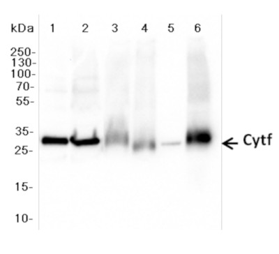 Anti-PetA antibodies (AS20 4377) Western blot result courtesy of Dr. Elena Pojidaeva Laboratory of Plant Gene Expression Timiryazev Institute of Plant Physiology RAS 127276 Moscow, Russia. More details of the applied Western blot protocol can be found here. Agrisera Western blot protocol example can be found here. Western blot video tutorials - watch here. Agrisera Western Blot Trouble Shooting Brochure, request here. |
Technical tips
Latest
Are blocking conditions nothing to pay attention to?2026-06-18 What are the most common misconceptions about primary antibodies?
2026-05-27 What are the most common mistakes done with secondary antibodies?
2026-05-26 Simple extraction protocol for membrane proteins
2026-04-17 How to improve signal with a weak antibody?
2026-03-26 Same antibody, different outcome on samples from wide taxonomic range
2026-02-23 Can the quality of protein transfer be checked before the blot is developed with antibodies?
2026-01-16 Is wild type a wild type?
2025-12-29 Why an antibody may detect tagged protein but not endogenous one and in some cases endogenous protein but not its tagged version?
2025-10-30 Calclulated and aparent molecular weight of detected protein is different, why?
2025-10-09
Archive
- June - 2026
- May - 2026
- April - 2026
- March - 2026
- February - 2026
- January - 2026
- December - 2025
- October - 2025
- September - 2025
- August - 2025
- July - 2025
- June - 2025
- May - 2025
- April - 2025
- March - 2025
- February - 2025
- January - 2025
- December - 2024
- November - 2024
- September - 2024
- July - 2024
- June - 2024
- May - 2024
- March - 2024
- February - 2024
- December - 2023
- November - 2023
- September - 2023
- July - 2023
- May - 2023
- March - 2023
- January - 2023
- December - 2022
- November - 2022
- October - 2022
- September - 2022
- August - 2022
- June - 2022
- May - 2022
- March - 2022
- February - 2022
- January - 2022
- November - 2021
- October - 2021
- August - 2021
- June - 2021
- May - 2021
- April - 2021
- March - 2021
- February - 2021
- January - 2021
- December - 2020
- November - 2020
- October - 2020
- September - 2020
- August - 2020
- July - 2020
- June - 2020
- May - 2020
- April - 2020
- March - 2020
- January - 2020
- November - 2019
- October - 2019
- March - 2019
- April - 2017
- February - 2017
- May - 2016
- February - 2014
- September - 2013
- December - 2010