Plant organelle/membrane isolation
- Arabidopsis lumen extraction
- Arabidopsis thylakoid extraction
- BBY preparation
- Chlorophyll measurements
- Intact chloroplast isolation method
- Mitochondrial fraction
- Nuclear fraction
- Plasma membrane fraction
- PSII RC extraction for cryo-EM
- Thylakoid extraction
- Vacuol isolation
- Collection of articles

- Diatom protein extraction
- Extraction of leaf proteins
- Phenol protein extraction
- Ponceau membrane staining
- Protein extraction from grasses
- TCA acetone precipitation method
- Western blot protocol
- Western blot video tutorial
- Western blot troubleshooting

- Western blot using IgY
- Western blot in denatured conditions (urea gel)

- Peptide neutralization/competition assay
- Quantitative Western blot
- Quantitative Western blot video tutorial
- Simultaneous Western blot
- Anti-KLH antibody removal

- Dot blot
- ELISA
- Immunohistochemistry
- Immunoprecipitation
- Immunoprecipitation/IgY
- Meiotic staining
- Yolk delipidation
Technical information
Antibody typesPurification
- Antibody purification
- Antibody purification - small amount of protein
- Elution of antibodies from affinity columns
- IgY purification methods
- Protein purification using antibodies
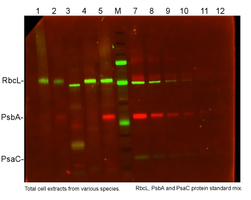
Protocols > Simultaneous Western blotSimultaneous Western blot using three different primary antibodies on the same blot.Procedure 1 μg of a total cell extract from (1) Arabidopsis thaliana, (2) Zea mays, (3) Anabaena sp., (4) Chlamydomonas reinhardtii, and (5) Thalassiosira pseudonana were separated on the Invitrogen NuPage system (Bis-Tris gel) for 50 min in MES buffer, then transferred to the Immobilon low FL 0.45 µm pvdf for 50 min. M - Biorad "WesternC" marker which did not cross-react with antibodies was used. Protein standard mix (7-12): RbcL (AS01 017S), PsbA (AS01 016S), PsaC (AS04 042S) in serial dilution 6X with a starting volume of 800 fmoles. Primary antibodies: rabbit anti-RbcL (AS03 037), chicken anti-PsbA (AS01 016) and rabbit anti-PsaC (AS10 939) were applied in 1: 2000 dilution followed by a standard washing protocol. The following secondary antibodies were used: goat anti-chicken DyLight® 488 conjugated (AS09 622) in dilution 1: 2000 (to detect PsbA) and goat anti-rabbit DyLight® 549 conjugated in dilution 1: 1000 (to detect RbcL and PsaC) followed by a standard washing protocol. The membrane was exposed for 3 s under the multiplex setting (DyLight ® 488 and DyLight ® 549) on the Biorad VersaDoc 4000 MP machine. Details on the excitation sources and emission filters of the VersaDoc can be found here. Note: primary antibodies to use in such a set up should be of equal titer. Chosen Agrisera secondary antibodiesAnti-chicken DyLight®-conjugated secondary antibodies AS11 1820 DyLight® 350 (violet) AS16 3264 DyLight® 405 (blue) AS09 622 DyLight® 488 (green) AS11 1822 DyLight® 550 (yellow) AS11 1823 DyLight® 594 (orange) AS11 1824 DyLight® 633(red) AS11 1825 DyLight® 650 (red-brown) AS11 1827 DyLight® 800 (near-IR) Anti-rabbit DyLight®-conjugated secondary antibodies AS11 1812 DyLight® 350 (violet) AS16 3510 DyLight® 405 (blue) AS16 3527 DyLight® 488 (green) AS11 1814 DyLight® 550 (yellow) AS11 1815 DyLight® 594 (orange) AS11 1816 DyLight® 633 (red) AS12 2331 DyLight® 650 (red-brown) AS16 3517 DyLight® 680 (far-red) AS12 2456 DyLight® 800 (near-IR) DyLight® is a trademark of Thermo Fisher Scientific, Inc. and its subsidiaries. |

