| Material: Zea mays hybrid variety Preparation 5 days-old germinating maize root tips were cut and fixed with 4% formaldehyde, 60 min, RT. Cell walls were digested with enzymes cellulase and pectinase in MES buffer for 90 min, RT. After washing with PBS, roots were squashed gently using a flat tip forceps to release the cells into PBS buffer. Cells were allowed to settle O/N at 4°C, followed by immunolocalization. Immunocytochemistry (ICC) Cells were permeabilized with 0.5% Triton X-100, 10 min, RT, followed by washing with PBS buffer before blocking with 5% fish gelatin-PBS, 30 min, RT. Detection antibody: Cells were incubated with rabbit anti-clathrin 1,2 primary antibodies conjugated with DyLight®594 (AS10 690-DL594, Agrisera) for 3h/RT. Nuclei were stained with DAPI followed Fluoromount-G mounting (Southern Biotech). Courtesy of Dr. Ferhan Ayaydin and Dr. Divya Teja Dondapati, Hungarian Centre of Excellence for Molecular Medicine, (HCEMM), Szeged, Hungary. | 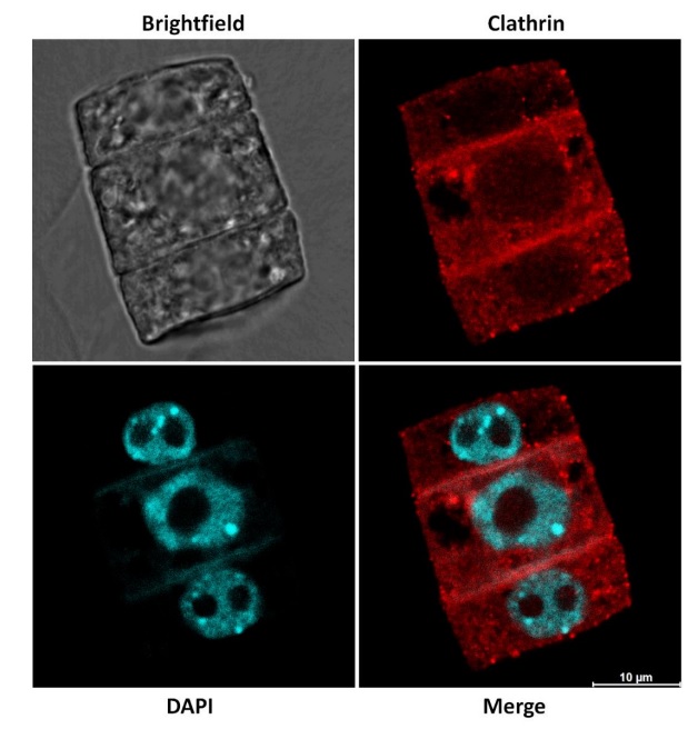 Upper panels show the Differential Interference Contrast (DIC) transmitted light image (gray) and Clathrin immunodetection (red). Lower panels represent DAPI labelled nuclei (cyan) and the merged image of Clathrin and DAPI. |
Agrisera News
Latest
Agrisera at the 36th International Conference on Arabidopsis Research 20262026-07-10 Agrisera supported five conferences in June
2026-06-26 Agrisera Team celebrates middle of the summer, Midsummer
2026-06-19 Agrisera Western blot workshop at BioLOCK conference in Poland
2026-06-08 Agrisera at the 7th International Plant Protease Conference 2026
2026-06-08 Innovation Day at Agrisera
2026-06-06 Anti-RbcL antibody used in several publications in 2026
2026-06-01 Agrisera supported three conferences in May
2026-05-29 Prof. Yoselin Benitez-Alfonso visited Agrisera facility in Umeå
2026-05-25 Agrisera antibodies used to show how plant derived photosynthetic neo-organelles can provide energy input into mammalian metabolic systems
2026-05-19
Archive
- July - 2026
- June - 2026
- May - 2026
- April - 2026
- March - 2026
- February - 2026
- January - 2026
- December - 2025
- November - 2025
- October - 2025
- September - 2025
- August - 2025
- July - 2025
- June - 2025
- May - 2025
- April - 2025
- March - 2025
- February - 2025
- January - 2025
- December - 2024
- November - 2024
- October - 2024
- September - 2024
- August - 2024
- July - 2024
- June - 2024
- May - 2024
- April - 2024
- March - 2024
- February - 2024
- January - 2024
- December - 2023
- November - 2023
- October - 2023
- September - 2023
- August - 2023
- July - 2023
- June - 2023
- May - 2023
- April - 2023
- March - 2023
- February - 2023
- January - 2023
- December - 2022
- November - 2022
- October - 2022
- September - 2022
- August - 2022
- July - 2022
- June - 2022
- May - 2022
- April - 2022
- March - 2022
- February - 2022
- January - 2022
- December - 2021
- November - 2021
- October - 2021
- September - 2021
- August - 2021
- July - 2021
- June - 2021
- May - 2021
- April - 2021
- March - 2021
- February - 2021
- January - 2021
- December - 2020
- November - 2020
- October - 2020
- September - 2020
- August - 2020
- July - 2020
- June - 2020
- May - 2020
- April - 2020
- March - 2020
- February - 2020
- January - 2020
- December - 2019
- November - 2019
- October - 2019
- September - 2019
- August - 2019
- July - 2019
- June - 2019
- May - 2019
- April - 2019
- March - 2019
- February - 2019
- January - 2019
- December - 2018
- November - 2018
- October - 2018
- September - 2018
- August - 2018
- July - 2018
- June - 2018
- May - 2018
- April - 2018
- March - 2018
- February - 2018
- January - 2018
- December - 2017
- November - 2017
- October - 2017
- September - 2017
- August - 2017
- July - 2017
- June - 2017
- April - 2017
- March - 2017
- February - 2017
- December - 2016
- November - 2016
- October - 2016
- September - 2016
- August - 2016
- July - 2016
- June - 2016
- May - 2016
- April - 2016
- March - 2016
- February - 2016
- January - 2016
- December - 2015
- November - 2015
- October - 2015
- September - 2015
- August - 2015
- July - 2015
- June - 2015
- May - 2015
- March - 2015
- February - 2015
- January - 2015
- December - 2014
- November - 2014
- October - 2014
- September - 2014
- August - 2014
- July - 2014
- June - 2014
- May - 2014
- April - 2014
- March - 2014
- February - 2014
- January - 2014
- December - 2013
- November - 2013
- September - 2013
- August - 2013
- July - 2013
- June - 2013
- May - 2013
- April - 2013
- February - 2013
- January - 2013
- December - 2012
- October - 2012
- September - 2012
- August - 2012
- July - 2012
- June - 2012
- May - 2012
- April - 2012
- March - 2012
- December - 2011
- November - 2011
- September - 2011
- August - 2011
- July - 2011
- April - 2011
- January - 2011
- December - 2010
- October - 2010
- September - 2010
- August - 2010
- July - 2010
- March - 2010
- January - 2010
- December - 2009
- November - 2009
- September - 2009
- July - 2009
- June - 2009
- May - 2009
- March - 2009
- January - 2009
- December - 2008